Tooth Anatomy Diagram Generator
Create a labeled tooth anatomy diagram with a clear longitudinal cutaway. Use the reference to distinguish hard tissues, the pulp space, root structures and the tissues that support the tooth. This is educational and not for diagnosis.
SEE ACADRAW IN ACTION
Science drawing, from idea to editable figure
Create a science illustration with AI. Refine drawings, build editable diagrams, and convert images to SVG.
Demos play as you scroll into view. Choose a feature to explore.
Trusted by students & researchers from 480+ universities
A global community spanning 60+ countries and regions, from QS top-ranked universities to leading research institutes
























University logos belong to their respective institutions and are shown solely to illustrate our user community. They do not imply any partnership or endorsement.
Tool overview
Explore the use cases, workflow, and key things to know.
Define the view
Choose a crown, root or full cutaway view before generating.
Keep tissue layers distinct
Use separate labels for enamel, dentin, pulp and supporting tissues.
Review before sharing
Check orientation and anatomy against reliable sources.
Labeled reference
Tooth anatomy reference: layers, roots and supporting tissues
The cutaway separates enamel, dentin, pulp, root canal, cementum, periodontal ligament, gum and jaw bone so learners can trace the tooth from crown to root. It is a simplified teaching illustration and is not a clinical image.
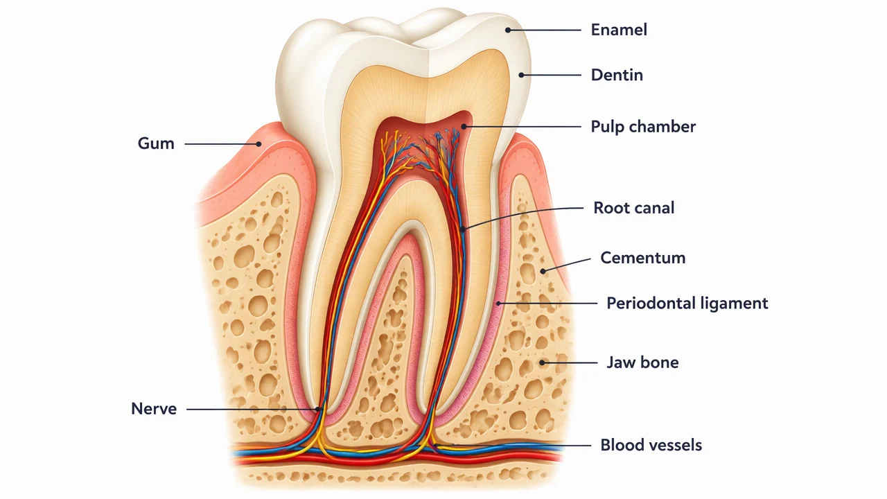
Tooth anatomy label key
Use this label key to plan your diagram. Soft tissues or functions described in the table may require an additional view or layer.
| Structure | Where to find it | Role and diagram note |
|---|---|---|
| Enamel | Outer crown surface | Hard protective tissue covering the visible crown. |
| Dentin | Below enamel and around the pulp | Forms most of the tooth and surrounds the internal pulp space. |
| Pulp chamber and root canal | Inside the crown and root | Continuous internal space containing vessels and nerves in the simplified model. |
| Cementum and periodontal ligament | Around the root | Attach and support the tooth within the surrounding tissues. |
| Gum and jaw bone | Around the tooth | Show the supporting context without implying a clinical examination. |
Anatomical relationships: OpenStax: The mouth, pharynx and esophagus. On small screens, scroll the table horizontally.
Read a tooth diagram from crown to root
Start at the crown, move through enamel and dentin to the pulp, then follow the root canal into the supporting tissues.
01
A cutaway selects one view
It reveals internal layers while omitting fine microscopic detail.
02
Hard and soft tissues differ
Keep enamel and dentin separate from pulp, gum and the periodontal ligament.
03
Orientation matters
Label crown, root and surrounding jaw context so readers know the direction.
Common tooth anatomy diagram mistakes
Mixing crown and root labels
Place crown structures above the gum and root structures below it.
Calling a diagram a diagnosis
Use it for education; symptoms require a qualified clinician.
Overloading microscopic detail
Add an inset when enamel prisms or cellular layers are the actual lesson.
Create your own tooth anatomy diagram
Start with the prefilled prompt in the generator above. Reading the reference is free; creating a new AI draft requires an account and available credits. See current pricing.
Choose Science Drawing for an illustrated draft, or Editable Diagram when you need separate shapes and text labels. Check the anatomy in either mode before using the result.
- 1
Choose the cutaway
Decide whether the lesson needs crown layers, root structures or both.
- 2
Group the labels
Keep hard tissues, pulp and supporting tissues visually distinct.
- 3
Review the anatomy
Check orientation and every leader-line endpoint against reliable anatomy references.
A prompt you can adapt
Create a clean, editable educational tooth anatomy cutaway with short readable labels for enamel, dentin, pulp chamber, root canal, gum, cementum, periodontal ligament, jaw bone, blood vessels and nerve. Use a longitudinal molar view, generous whitespace and a note that the illustration is simplified and not for diagnosis.
Use this prompt in the generatorTooth Anatomy FAQ
What are the main parts of a tooth?
An introductory cutaway commonly labels enamel, dentin, pulp, root canal, cementum, gum and supporting tissues.
What is the difference between enamel and dentin?
Enamel covers the crown; dentin lies beneath it and forms much of the tooth.
Where is the pulp?
The pulp occupies the pulp chamber and continues through the root canal in the simplified model.
Can this diagram diagnose a dental problem?
No. It is an educational illustration and cannot replace a dental examination.
Can I make a blank tooth worksheet?
Yes. Keep the checked labeled version as an answer key and remove labels from a duplicate.
Sources and editorial notes
- OpenStax, Anatomy and Physiology: The mouth, pharynx and esophagus — factual background for the accompanying explanation.
- OpenStax: The Stomach and Digestive System — additional educational context.
Prepared by the AcaDraw product team. Source and label check: . Independent medical review has not been completed. This material is for education; AI drafts need review against reliable anatomy references before use and are not for diagnosis or treatment.
Turn the reference into your own teaching figure
Choose the structures your lesson needs, create a draft, and review it before sharing.
Read the tooth anatomy guideSkin Diagram
Explore the labeled reference and drawing guide.
Urinary System Diagram
Explore the labeled reference and drawing guide.
Endocrine System Diagram
Explore the labeled reference and drawing guide.
Eye Anatomy Diagram
Explore the labeled reference and drawing guide.
Human Anatomy Diagram
Explore the labeled reference and drawing guide.
Ear Anatomy Diagram
Explore the labeled reference and drawing guide.
Lymphatic System Diagram
Explore the labeled reference and drawing guide.
Muscular System Diagram
Explore the labeled reference and drawing guide.
Kidney diagram
Explore the labeled reference and drawing guide.
Nephron diagram
Explore the labeled reference and drawing guide.
Stomach diagram
Explore the labeled reference and drawing guide.
Female reproductive system diagram
Explore the labeled reference and drawing guide.
Lungs diagram
Explore the labeled reference and drawing guide.
Nervous system diagram
Explore the labeled reference and drawing guide.
Labeled heart diagram
Explore the labeled reference and drawing guide.
Foot Anatomy diagram
Explore the labeled reference and drawing guide.
Shoulder Anatomy diagram
Explore the labeled reference and drawing guide.
Labeled brain diagram
Explore the labeled reference and drawing guide.
Digestive system diagram
Explore the labeled reference and drawing guide.
Respiratory system diagram
Explore the labeled reference and drawing guide.
Skeletal system diagram
Explore the labeled reference and drawing guide.
Neuron diagram
Explore the labeled reference and drawing guide.
Brainstem
Explore the labeled reference and drawing guide.
Medical illustration generator
Plan other educational anatomy illustrations.