Shoulder Anatomy Diagram Generator
Understand shoulder anatomy with a labeled joint reference, the three main bones, and a guide to the rotator cuff. Create a study diagram with the right view and layers.
SEE ACADRAW IN ACTION
Science drawing, from idea to editable figure
Create a science illustration with AI. Refine drawings, build editable diagrams, and convert images to SVG.
Demos play as you scroll into view. Choose a feature to explore.
Trusted by students & researchers from 480+ universities
A global community spanning 60+ countries and regions, from QS top-ranked universities to leading research institutes





















































































University logos belong to their respective institutions and are shown solely to illustrate our user community. They do not imply any partnership or endorsement.
Tool overview
Explore the use cases, workflow, and key things to know.
Three main bones
Locate the clavicle, scapula and humerus before adding soft tissue.
Four cuff muscles
Use SITS to remember supraspinatus, infraspinatus, teres minor and subscapularis.
View-aware labels
Use separate anterior, posterior and sectional views rather than exposing every structure at once.
Labeled reference
Start with the ball, socket and supporting tissues
This section through the shoulder reveals the humeral head, glenoid cavity, labrum, capsule and nearby tendons. It is a sectional joint reference, not an external view of all four rotator cuff muscles.
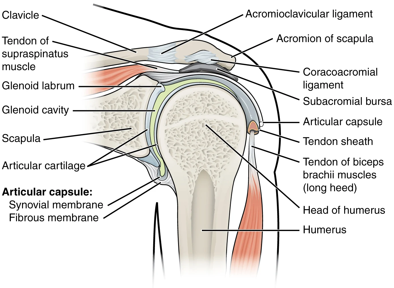
Shoulder anatomy labels and relationships
Use this label key alongside the reference. Soft tissues or functions described in the table may require an additional view or layer.
| Structure | Where it belongs | Function or drawing note |
|---|---|---|
| Clavicle | Between sternum and acromion | Connects the upper limb girdle to the axial skeleton through the sternoclavicular joint. |
| Scapula and glenoid cavity | Shoulder blade and its lateral socket | The shallow glenoid receives the humeral head. The acromion extends over the joint. |
| Humeral head | Proximal humerus | The ball of the glenohumeral joint. Do not confuse it with the acromioclavicular joint above. |
| Glenoid labrum | Rim around the glenoid | Fibrocartilaginous rim that deepens the socket; distinct from the smooth articular cartilage. |
| Capsule and glenohumeral ligaments | Around the glenohumeral joint | Provide passive restraint while allowing a wide range of motion. |
| Supraspinatus | Above the scapular spine, tendon passes beneath the acromion | Assists initiation of abduction and stabilizes the humeral head. |
| Infraspinatus and teres minor | Posterior scapula | Externally rotate the arm and contribute to humeral-head stability. |
| Subscapularis | Anterior surface of the scapula | Internally rotates the arm and contributes to joint stability; not fully visible on a posterior view. |
| Deltoid, long-head biceps tendon and bursa | Superficial shoulder, near the joint and below acromion | Deltoid is not a cuff muscle. Biceps tendon and subacromial bursa are adjacent structures, not extra cuff muscles. |
Anatomical relationships: OpenStax: Anatomy of selected synovial joints. On small screens, scroll the table horizontally.
The shoulder complex is more than one joint
Use “shoulder joint” precisely when a drawing concerns the glenohumeral articulation. Raising the arm also depends on movement of the shoulder girdle.
01
Glenohumeral joint: ball and socket
The humeral head articulates with the glenoid cavity. A shallow socket permits mobility; the labrum, capsule, ligaments and muscle activity contribute to stability.
02
Acromioclavicular and sternoclavicular joints
The AC joint joins the clavicle to the acromion. The SC joint joins the clavicle to the sternum. Do not label the AC joint as the socket for the humeral head.
03
Scapulothoracic movement and the rotator cuff
The scapula glides over the thoracic wall as a functional articulation, not a typical synovial joint. The four cuff muscles help center the humeral head during movement; show their positions in separate anterior and posterior panels.
Common shoulder anatomy diagram mistakes
Adding the deltoid to the rotator cuff
The four cuff muscles are SITS. The deltoid is an important mover of the arm, but it belongs to a different muscle group.
Drawing all cuff muscles on one surface
Subscapularis is anterior to the scapula; infraspinatus and teres minor are posterior. Use multiple views or clearly explained cutaways.
Confusing cartilage, labrum and bursa
Articular cartilage covers joint surfaces, the labrum rims the glenoid, and a bursa reduces friction between adjacent tissues. They are separate structures.
Create your own shoulder anatomy diagram
Start with the prefilled prompt in the generator above. Reading the reference is free; creating a new AI draft requires an account and available credits. See current pricing.
Choose Science Drawing for an illustrated draft, or Editable Diagram when you need separate shapes and text labels. Check the anatomy in either mode before using the result.
- 1
Choose the anatomical question
Use a section for joint surfaces and capsule. Use anterior and posterior views for the four rotator cuff muscles.
- 2
Generate separate panels
Specify the side, view, visible tissues and exact label list. Avoid combining deep and superficial layers without a cutaway key.
- 3
Verify the layers and attachments
Check cuff membership, humeral-head position and each leader line against a reference. Edit the draft before sharing or exporting.
A prompt you can adapt
Create a clean educational shoulder anatomy illustration with three panels: sectional glenohumeral joint, anterior rotator cuff, posterior rotator cuff. Label anatomical side and view. In the section label clavicle, acromion, scapula, glenoid cavity, labrum, humeral head, capsule, supraspinatus tendon and subacromial bursa. In the muscle views label supraspinatus, infraspinatus, teres minor and subscapularis in their correct locations. Distinguish deltoid from the cuff. Use spaced editable labels and leader lines on white. Do not invent tears, diagnoses or surgical findings.
Use this prompt in the generatorShoulder Anatomy FAQ
What are the three main bones of the shoulder?
The clavicle, scapula and humerus. The clavicle and scapula form the pectoral girdle; the proximal humerus participates in the shoulder joint.
Which muscles form the rotator cuff?
Supraspinatus, infraspinatus, teres minor and subscapularis. SITS is a common memory aid. Deltoid and teres major are not cuff muscles.
What is the difference between the AC and shoulder joints?
The acromioclavicular joint is between the clavicle and acromion. The glenohumeral joint is between the humeral head and glenoid cavity.
Where is the labrum in a shoulder diagram?
It lies around the rim of the glenoid cavity and deepens the socket. It is separate from the articular cartilage covering the joint surfaces.
Can I use this diagram to identify a rotator cuff tear?
No. This is an educational anatomy reference. Diagnosing a tear requires clinical assessment and may require imaging; an AI illustration is not diagnostic evidence.
How do I create a labeled shoulder diagram?
Use the prompt above to start a draft in AcaDraw, choose the views and inspect every structure against the cited references. Sign-in and available credits are required for generation.
Sources and editorial notes
- OpenStax, Anatomy and Physiology: Anatomy of selected synovial joints — primary anatomy reference and illustration source.
- AAOS OrthoInfo: Rotator Cuff Tears — additional educational context.
Prepared by the AcaDraw product team. Source and label check: . Independent medical review has not been completed. This material is for education; AI drafts need review against reliable anatomy references before use and are not for diagnosis or treatment.
Turn the reference into your own teaching figure
Choose the structures your lesson needs, create a draft, and review it before sharing.
Read the step-by-step tutorial