Lymphatic System Diagram Generator
Create a lymphatic system diagram that distinguishes vessels, node groups and lymphoid organs. Use an anterior body overview to orient the spleen and major drainage routes, then add a separate inset when the lesson needs node structure.
SEE ACADRAW IN ACTION
Science drawing, from idea to editable figure
Create a science illustration with AI. Refine drawings, build editable diagrams, and convert images to SVG.
Demos play as you scroll into view. Choose a feature to explore.
Trusted by students & researchers from 480+ universities
A global community spanning 60+ countries and regions, from QS top-ranked universities to leading research institutes
























University logos belong to their respective institutions and are shown solely to illustrate our user community. They do not imply any partnership or endorsement.
Tool overview
Explore the use cases, workflow, and key things to know.
Orient the lymphatic system diagram
Use patient-left labels before identifying the spleen and thoracic duct return.
Distinguish nodes from organs
Lymph nodes filter lymph; the spleen filters blood and the thymus supports T-cell maturation.
Trace return toward veins
Lymph rejoins venous circulation rather than circulating through a second heart-driven loop.
Labeled reference
Lymphatic system diagram: vessels, nodes, and lymphoid organs
This lymphatic system diagram is a simplified anterior overview. Patient left appears on the viewer’s right. Green paths illustrate vessel distribution rather than every real vessel. Node clusters are schematic; the spleen lies in the patient’s left upper abdomen and filters blood rather than lymph.
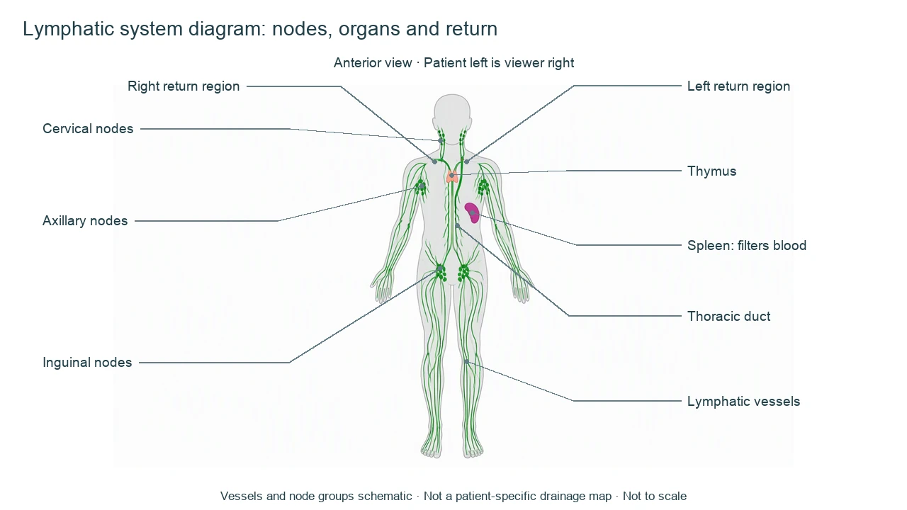
Lymphatic system diagram label key
Use this label key to plan your diagram. Soft tissues or functions described in the table may require an additional view or layer.
| Label or process | Position or relationship | Meaning and drawing check |
|---|---|---|
| Lymphatic capillaries and vessels | Distributed through many tissues | Return excess interstitial fluid and proteins toward venous blood; the overview does not map every vessel. |
| Cervical nodes | Neck region | One regional node group; exact number and location vary and are simplified. |
| Axillary nodes | Armpit region | Receive lymph from territories including the upper limb and parts of the thorax; drainage is more detailed than this overview. |
| Inguinal nodes | Groin region | Receive lymph from several lower-body territories; not the only lower-limb nodes. |
| Thymus | Upper anterior mediastinum | Supports T-cell maturation; appearance changes with age. |
| Spleen | Patient's left upper abdomen | Filters blood and supports immune responses. It is not a large lymph node in the vessel chain. |
| Thoracic duct | Major central lymphatic conduit | Drains most of the body toward the left venous angle, with variations in anatomy. |
| Right lymphatic drainage | Right upper quadrant in the conventional model | Returns lymph from the right side of head/neck, right upper limb and right thorax; other territories usually drain through the thoracic duct. |
Anatomical relationships: OpenStax: Anatomy of the Lymphatic and Immune Systems. On small screens, scroll the table horizontally.
Follow a lymphatic system diagram from tissues toward venous return
A lymphatic system diagram combines fluid return, immune tissues and specialized intestinal absorption. These functions need distinct explanations rather than a single unlabeled green line.
01
Fluid return has a direction
Interstitial fluid enters lymphatic capillaries and travels through vessels and nodes before returning to veins. Valves and external movements help flow; there is no lymphatic heart.
02
Nodes and spleen encounter different fluids
Lymph nodes sample lymph from tissues, while the spleen monitors blood. Do not draw the spleen as a mandatory filter along every lymph vessel.
03
Intestinal lacteals need an inset
Lacteals help absorb dietary lipids from the small intestine. This body overview omits villus-level detail and does not show a complete nutrient transport map.
Common lymphatic system diagram mistakes
Putting the spleen on patient right
In this anterior lymphatic system diagram, the spleen is on viewer right, corresponding to patient left.
Saying all lymph drains to the right duct
The thoracic duct serves most territories in the conventional model; the right upper quadrant has distinct return.
Treating the schematic as a clinical map
A simplified lymphatic system diagram cannot diagnose swollen nodes or identify an individual's drainage pattern.
Create your own lymphatic system diagram
Start with the prefilled prompt in the generator above. Reading the reference is free; creating a new AI draft requires an account and available credits. See current pricing.
Choose Science Drawing for an illustrated draft, or Editable Diagram when you need separate shapes and text labels. Check the anatomy in either mode before using the result.
- 1
Choose the view
Request an anterior lymphatic system diagram and state patient left/right explicitly.
- 2
Separate label layers
List vessels, regional nodes and lymphoid organs without merging their functions.
- 3
Check return and laterality
Review the spleen side and the two venous return regions before sharing.
A prompt you can adapt
Create a labeled anterior lymphatic system diagram of a gender-neutral adult. Mark patient LEFT on viewer right. Show schematic green lymph vessels and cervical, axillary and inguinal nodes, thymus in upper chest, spleen in left upper abdomen, central thoracic duct returning near the left venous angle and conventional right upper-quadrant lymphatic drainage toward the right venous angle. Distinguish lymph-filtering nodes from blood-filtering spleen. Do not invent exact vessel anatomy, clinical findings or flow rates. White background, noncrossing leaders, simplified and not to scale.
Use this prompt in the generatorLymphatic system FAQ
What should a lymphatic system diagram include?
A basic lymphatic system diagram includes vessels, selected node groups, thymus, spleen and major drainage routes. A detailed lesson may add tonsils, intestinal lymphoid tissue or lacteals.
Where is the spleen in an anterior view?
It is in the patient's left upper abdomen, which appears on the viewer's right. The spleen filters blood rather than lymph.
What is the difference between lymph and interstitial fluid?
Fluid between tissue cells is interstitial fluid. After entering lymphatic capillaries it is called lymph and is returned toward venous blood.
Where does the thoracic duct drain?
In the conventional model it returns lymph from most of the body toward the left venous angle near the junction of the internal jugular and subclavian veins; anatomical variation exists.
Does lymph flow in a closed loop like blood?
No. Lymphatic capillaries collect fluid from tissues and return it to venous circulation. Flow depends on valves and pressure changes rather than a separate central pump.
Can this diagram diagnose a swollen node?
No. This is a simplified educational model, not a clinical assessment or patient-specific map.
Can I create an editable lymphatic system diagram?
Yes. Choose Editable Diagram for shapes and labels or Science Drawing for an illustration. Generation requires an account and credits; review the anatomy.
Sources and editorial notes
- OpenStax, Anatomy and Physiology: Anatomy of the Lymphatic and Immune Systems — factual background for the accompanying explanation.
- OpenStax: Barrier Defenses and the Innate Immune Response — additional educational context.
Prepared by the AcaDraw product team. Source and label check: . Independent medical review has not been completed. This material is for education; AI drafts need review against reliable anatomy references before use and are not for diagnosis or treatment.
Turn the reference into your own teaching figure
Choose the structures your lesson needs, create a draft, and review it before sharing.
Read the lymphatic system anatomy guideMuscular System Diagram
Explore the labeled reference and drawing guide.
Kidney diagram
Explore the labeled reference and drawing guide.
Nephron diagram
Explore the labeled reference and drawing guide.
Stomach diagram
Explore the labeled reference and drawing guide.
Female reproductive system diagram
Explore the labeled reference and drawing guide.
Lungs diagram
Explore the labeled reference and drawing guide.
Nervous system diagram
Explore the labeled reference and drawing guide.
Labeled heart diagram
Explore the labeled reference and drawing guide.
Foot Anatomy diagram
Explore the labeled reference and drawing guide.
Shoulder Anatomy diagram
Explore the labeled reference and drawing guide.
Labeled brain diagram
Explore the labeled reference and drawing guide.
Digestive system diagram
Explore the labeled reference and drawing guide.
Respiratory system diagram
Explore the labeled reference and drawing guide.
Skeletal system diagram
Explore the labeled reference and drawing guide.
Neuron diagram
Explore the labeled reference and drawing guide.
Medical illustration generator
Plan other educational anatomy illustrations.