Muscular System Diagram Generator
Create a muscular system diagram that separates anterior and posterior views. Label major superficial skeletal muscles, distinguish individual muscles from muscle groups, and choose a focused layer before adding movement arrows.
SEE ACADRAW IN ACTION
Science drawing, from idea to editable figure
Create a science illustration with AI. Refine drawings, build editable diagrams, and convert images to SVG.
Demos play as you scroll into view. Choose a feature to explore.
Trusted by students & researchers from 480+ universities
A global community spanning 60+ countries and regions, from QS top-ranked universities to leading research institutes
























University logos belong to their respective institutions and are shown solely to illustrate our user community. They do not imply any partnership or endorsement.
Tool overview
Explore the use cases, workflow, and key things to know.
Choose a view and a layer
A muscular system diagram should say anterior, posterior and superficial before naming the visible muscles.
Separate muscles from groups
Quadriceps and hamstrings are groups; biceps brachii and rectus abdominis are individual muscles.
Review movement context
Muscle action depends on joints, position and cooperation; a static overview is not a complete biomechanics model.
Labeled reference
Muscular system diagram: major superficial muscles, front and back
This muscular system diagram is a simplified adult overview of selected superficial skeletal muscles. Anterior and posterior figures show different surfaces; they do not reveal every deep muscle. Group labels such as quadriceps and hamstrings represent several muscles, and the illustration does not encode strength or injury.
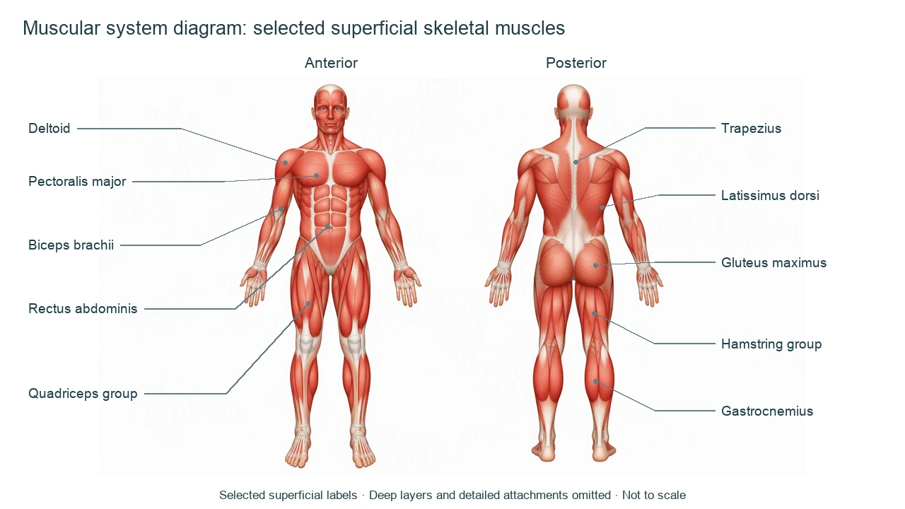
Muscular system diagram labels and movement context
Use this label key to plan your diagram. Soft tissues or functions described in the table may require an additional view or layer.
| Label or process | Position or relationship | Meaning and drawing check |
|---|---|---|
| Deltoid | Superficial shoulder | Different portions contribute to different shoulder movements; do not give every fiber one identical action. |
| Pectoralis major | Anterior chest | Contributes to humeral adduction and medial rotation, with part-specific actions. |
| Biceps brachii | Anterior upper arm | Supports forearm supination and elbow flexion; it is not the only elbow flexor. |
| Rectus abdominis | Paired anterior abdominal muscle | Contributes to trunk flexion and abdominal compression; visible segments do not represent separate muscles. |
| Quadriceps femoris group | Anterior thigh | Four muscles contribute to knee extension; rectus femoris also crosses the hip. |
| Trapezius | Superficial upper back and neck | Different fiber regions move and stabilize the scapula in different ways. |
| Latissimus dorsi | Broad posterior trunk | Contributes to extension, adduction and medial rotation of the humerus. |
| Gluteus maximus | Superficial buttock region | A major hip extensor; not the entire gluteal muscle group. |
| Hamstring group | Posterior thigh | Generally supports knee flexion and hip extension, with member-specific differences. |
| Gastrocnemius | Superficial posterior calf | Crosses knee and ankle, contributes to plantar flexion and knee flexion. |
Anatomical relationships: OpenStax: Axial Muscles of the Abdominal Wall, and Thorax. On small screens, scroll the table horizontally.
Read a muscular system diagram by region and function
A muscular system diagram can guide identification, but detailed actions require a joint and position. Use separate regional figures for attachments or deep structures.
01
Anterior and posterior are complementary views
Front and back labels refer to the same body, not alternative versions with interchangeable visible muscles.
02
Groups need explicit names
Quadriceps includes four muscles. Hamstrings commonly refers to a posterior-thigh group rather than a single large sheet.
03
Skeletal muscle is only one muscle tissue type
This overview does not map cardiac muscle in the heart or smooth muscle in organ walls; those need different diagrams.
Common muscular system diagram mistakes
Labeling a whole group as one muscle
State quadriceps group or hamstring group and use a regional inset when individual members matter.
Placing every muscle in the same surface view
Deep muscles are obscured by superficial ones; a front/back overview cannot show all layers at once.
Using the drawing for injury diagnosis
A muscular system diagram is an educational reference, not a diagnosis, rehabilitation plan or strength measurement.
Create your own muscular system diagram
Start with the prefilled prompt in the generator above. Reading the reference is free; creating a new AI draft requires an account and available credits. See current pricing.
Choose Science Drawing for an illustrated draft, or Editable Diagram when you need separate shapes and text labels. Check the anatomy in either mode before using the result.
- 1
Choose front, back or a region
Request a muscular system diagram with an explicit view and superficial or deep layer.
- 2
Set a manageable label list
Start with major visible muscles and identify group labels clearly.
- 3
Review laterality and actions
Check leader endpoints and qualify movements by joint before using the draft.
A prompt you can adapt
Create a labeled muscular system diagram with two gender-neutral adult figures: anterior view and posterior view. Show selected superficial skeletal muscles. Anterior labels: deltoid, pectoralis major, biceps brachii, rectus abdominis and quadriceps group. Posterior labels: trapezius, latissimus dorsi, gluteus maximus, hamstring group and gastrocnemius. Keep deep muscles out of the surface layer, distinguish groups from individual muscles and avoid duplicate leader endpoints. White background, readable noncrossing labels, no injury findings or quantitative strength claims, simplified and not to scale.
Use this prompt in the generatorMuscular system FAQ
What does a muscular system diagram show?
An introductory muscular system diagram usually shows major superficial skeletal muscles in anterior and posterior views, with labels for selected individual muscles and groups.
Does it show all muscles in the body?
No. Deep layers, small muscles and detailed attachments require additional regional views. This figure is a selected overview.
Are quadriceps and hamstrings single muscles?
No. They are muscle groups. A group label is useful in an overview, while detailed lessons should name the individual members.
Does the diagram include smooth and cardiac muscle?
This overview focuses on skeletal muscle. Smooth and cardiac muscle are different tissue types with different locations and structures.
Can one muscle have more than one action?
Yes. Actions depend on the joints crossed, fiber region and body position. Muscles also act together to stabilize and move joints.
Can I use it to diagnose pain or injury?
No. A simplified muscular system diagram is for education and cannot replace a clinical assessment.
Can I create an editable muscular system diagram?
Yes. Use Editable Diagram for separate shapes and labels or Science Drawing for an illustration. Generation requires an account and credits; review all anatomy.
Sources and editorial notes
- OpenStax, Anatomy and Physiology: Axial Muscles of the Abdominal Wall, and Thorax — factual background for the accompanying explanation.
- OpenStax: Muscles of the Pectoral Girdle and Upper Limbs — additional educational context.
Prepared by the AcaDraw product team. Source and label check: . Independent medical review has not been completed. This material is for education; AI drafts need review against reliable anatomy references before use and are not for diagnosis or treatment.
Turn the reference into your own teaching figure
Choose the structures your lesson needs, create a draft, and review it before sharing.
Read the muscular system anatomy guideSkin Diagram
Explore the labeled reference and drawing guide.
Urinary System Diagram
Explore the labeled reference and drawing guide.
Endocrine System Diagram
Explore the labeled reference and drawing guide.
Lymphatic System Diagram
Explore the labeled reference and drawing guide.
Kidney diagram
Explore the labeled reference and drawing guide.
Nephron diagram
Explore the labeled reference and drawing guide.
Stomach diagram
Explore the labeled reference and drawing guide.
Female reproductive system diagram
Explore the labeled reference and drawing guide.
Lungs diagram
Explore the labeled reference and drawing guide.
Nervous system diagram
Explore the labeled reference and drawing guide.
Labeled heart diagram
Explore the labeled reference and drawing guide.
Foot Anatomy diagram
Explore the labeled reference and drawing guide.
Shoulder Anatomy diagram
Explore the labeled reference and drawing guide.
Labeled brain diagram
Explore the labeled reference and drawing guide.
Digestive system diagram
Explore the labeled reference and drawing guide.
Respiratory system diagram
Explore the labeled reference and drawing guide.
Skeletal system diagram
Explore the labeled reference and drawing guide.
Neuron diagram
Explore the labeled reference and drawing guide.
Medical illustration generator
Plan other educational anatomy illustrations.