Neuron Diagram Generator
Read this labeled neuron diagram from dendrites and cell body to axon terminals. Locate myelin and nodes, distinguish electrical signaling from communication across a chemical synapse, then create a focused teaching draft.
SEE ACADRAW IN ACTION
Science drawing, from idea to editable figure
Create a science illustration with AI. Refine drawings, build editable diagrams, and convert images to SVG.
Demos play as you scroll into view. Choose a feature to explore.
Trusted by students & researchers from 480+ universities
A global community spanning 60+ countries and regions, from QS top-ranked universities to leading research institutes
























University logos belong to their respective institutions and are shown solely to illustrate our user community. They do not imply any partnership or endorsement.
Tool overview
Explore the use cases, workflow, and key things to know.
Find the main neuron parts
Separate dendrites, soma and axon before adding finer labels.
Trace signal direction
Use distinct annotations for input, an axonal action potential and synaptic transmission.
Make the teaching model explicit
Specify a multipolar neuron and CNS or PNS myelination rather than mixing cell types.
Labeled reference
Labeled neuron diagram: identify the parts before tracing signals
This AI-generated neuron diagram shows a typical multipolar cell with branching dendrites, soma, nucleus and a myelinated axon. Labels identify the major parts; the companion article explains signal direction and chemical synapses separately. Myelinating glial-cell bodies are omitted. Oligodendrocytes form CNS myelin, while Schwann cells form PNS myelin.
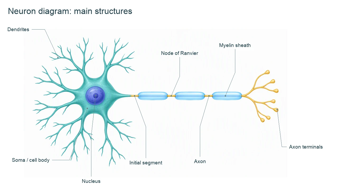
Neuron diagram labels and functions
Use this label key to plan your diagram. Soft tissues or functions described in the table may require an additional view or layer.
| Part | Location | Role and diagram note |
|---|---|---|
| Dendrites | Branching processes from the soma | Receive many synaptic inputs. Dendritic shape and function vary among neuron types. |
| Soma / cell body | Region around the nucleus | Contains organelles and supports the cell’s metabolism; inputs are integrated across the dendritic and somatic membrane. |
| Nucleus | Within the soma | Contains DNA. It is not where an axonal action potential starts. |
| Axon hillock and initial segment | Transition from soma into axon | For the typical model shown in teaching diagrams, an action potential usually initiates in the initial segment when threshold is reached. |
| Axon | Long process extending from the soma | Conducts action potentials toward terminals in the standard signaling model. An axon may have collateral branches. |
| Myelin sheath | Glial wrapping around axon segments | Insulates membrane and supports faster saltatory conduction. It is not one continuous sleeve or part of the axoplasm. |
| Nodes of Ranvier | Gaps between adjacent myelin segments | Sites where action potentials are regenerated in a myelinated axon. |
| Axon terminals | Branches at the far end of the axon | Presynaptic endings can release neurotransmitter at chemical synapses. |
| Synapse | Junction between communicating cells | A chemical synapse includes a presynaptic terminal, cleft and postsynaptic membrane. The target may be another neuron, muscle or gland. |
Anatomical relationships: OpenStax: Nervous tissue. On small screens, scroll the table horizontally.
Follow the signal through a neuron diagram
Use a neuron diagram to distinguish receiving input, conducting an action potential and transmitting a chemical signal. A single undifferentiated arrow can hide these changes.
01
Input and integration
Synaptic inputs commonly reach dendrites and the soma. Graded signals affect whether the initial segment reaches threshold. Not all inputs are excitatory.
02
Axonal conduction
An action potential is regenerated along the axonal membrane. In myelinated axons, regeneration occurs at nodes; saltatory conduction does not mean neurotransmitter jumps between nodes.
03
Chemical synaptic transmission
Arrival at a terminal triggers events that can release neurotransmitter. The transmitter crosses the cleft and binds receptors on the target; the same electrical impulse does not literally leap across the gap.
Common neuron diagram mistakes
Pointing the action-potential arrow at the nucleus
For the typical multipolar model, start the axonal action-potential arrow near the initial segment and point toward terminals.
Drawing myelin as a continuous tube
Leave nodes between segments. Distinguish the sheath from the axon inside it.
Mixing CNS and PNS myelination
Glial-cell bodies are omitted from this overview. If you add them, use oligodendrocytes for CNS myelin or Schwann cells for PNS myelin and name the context explicitly.
Create your own neuron diagram
Start with the prefilled prompt in the generator above. Reading the reference is free; creating a new AI draft requires an account and available credits. See current pricing.
Choose Science Drawing for an illustrated draft, or Editable Diagram when you need separate shapes and text labels. Check the anatomy in either mode before using the result.
- 1
Choose the neuron model
Start with a typical multipolar neuron; decide whether the lesson concerns CNS or PNS myelin.
- 2
Separate structure and signaling
Request readable labels plus a distinct action-potential arrow. Use a synaptic inset for the chemical step.
- 3
Verify before export
Check the initial segment, myelin gaps and transmitter direction. Keep drawing conventions separate from biological exceptions.
A prompt you can adapt
Create an educational labeled neuron diagram of a typical multipolar neuron. Label dendrites, soma, nucleus, axon hillock, initial segment, axon, myelin sheath, nodes of Ranvier and axon terminals. Show input arrows toward dendrites and soma, then a distinct action-potential arrow along the axon toward terminals. Add an enlarged chemical synapse inset showing transmitter release, cleft and postsynaptic receptors. Specify CNS myelin from oligodendrocytes; do not combine it with a Schwann-cell model. Make the overview and inset scale difference explicit. Review labels and signal directions.
Use this prompt in the generatorNeuron FAQ
What are the main parts of a neuron diagram?
A basic neuron diagram labels dendrites, soma, nucleus, axon, myelin sheath, nodes of Ranvier and axon terminals. Add an initial segment and a synapse inset when explaining signaling.
Which direction does a signal travel in a typical neuron diagram?
The introductory model shows inputs reaching dendrites and soma, an action potential starting near the initial segment, and axonal conduction toward terminals. This is a useful model, not a rule for every neuron type or experimental condition.
Are the axon terminal and synapse the same thing?
No. The terminal is the presynaptic ending. A chemical synapse includes that ending, a synaptic cleft and the postsynaptic membrane.
What is a node of Ranvier?
It is a gap in myelin along an axon where membrane channels support regeneration of the action potential. Myelin is segmented.
Do all neurons look like this diagram?
No. Multipolar, bipolar and pseudounipolar neurons have different arrangements. Not all axons are myelinated. Name the type and context shown.
Can I create an editable neuron diagram?
Yes. Use Editable Diagram for separate shapes and labels, or Science Drawing for an illustrated draft. An account and credits are required for generation, and the result needs a scientific review.
Sources and editorial notes
- OpenStax, Anatomy and Physiology: Nervous tissue — factual background for the accompanying explanation.
- OpenStax: Communication Between Neurons — additional educational context.
Prepared by the AcaDraw product team. Source and label check: . Independent medical review has not been completed. This material is for education; AI drafts need review against reliable anatomy references before use and are not for diagnosis or treatment.
Turn the reference into your own teaching figure
Choose the structures your lesson needs, create a draft, and review it before sharing.
Read the neuron parts and signal-direction guideLabeled heart diagram
Explore the labeled reference and drawing guide.
Foot Anatomy diagram
Explore the labeled reference and drawing guide.
Shoulder Anatomy diagram
Explore the labeled reference and drawing guide.
Labeled brain diagram
Explore the labeled reference and drawing guide.
Digestive system diagram
Explore the labeled reference and drawing guide.
Respiratory system diagram
Explore the labeled reference and drawing guide.
Skeletal system diagram
Explore the labeled reference and drawing guide.
Medical illustration generator
Plan other educational anatomy illustrations.