Skin Diagram Generator
Create a skin diagram that separates the epidermis and dermis from the underlying hypodermis. Locate a hair follicle, glands and a deep sensory receptor in a simplified section of thin, hairy skin.
SEE ACADRAW IN ACTION
Science drawing, from idea to editable figure
Create a science illustration with AI. Refine drawings, build editable diagrams, and convert images to SVG.
Demos play as you scroll into view. Choose a feature to explore.
Trusted by students & researchers from 480+ universities
A global community spanning 60+ countries and regions, from QS top-ranked universities to leading research institutes
























University logos belong to their respective institutions and are shown solely to illustrate our user community. They do not imply any partnership or endorsement.
Tool overview
Explore the use cases, workflow, and key things to know.
Two skin layers
Distinguish epidermis and dermis from the hypodermis below.
Hair and gland labels
Use a thin-skin section to locate follicles, ducts and selected receptors.
Teaching draft or worksheet
Review labels, then prepare a numbered skin diagram and answer key.
Labeled reference
Skin Diagram: labeled teaching reference
This skin diagram is a simplified section of thin, hairy skin. The epidermis is avascular; vessels are shown in deeper tissue. Hypodermis lies beneath skin proper. The section does not represent thick palmar skin or every receptor type.
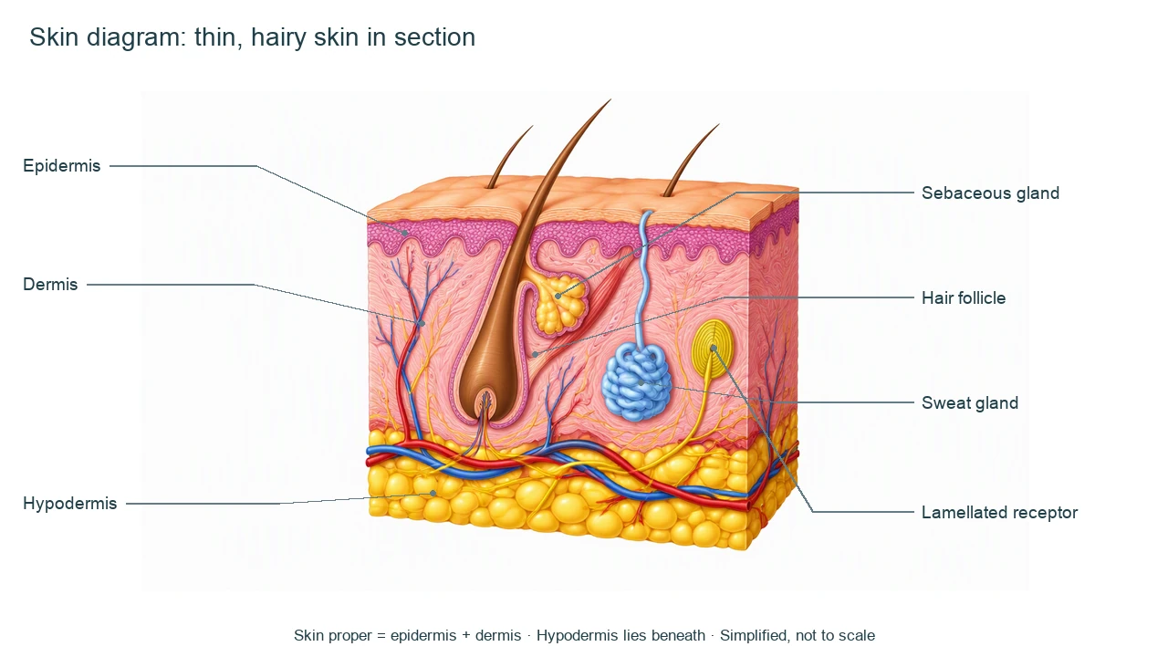
Skin Diagram label key
Use this label key to plan your diagram. Soft tissues or functions described in the table may require an additional view or layer.
| Label or stage | Position or relationship | Meaning and drawing check |
|---|---|---|
| Epidermis | Outer epithelial region | Avascular, keratinized stratified squamous epithelium. Do not place large blood vessels here. |
| Dermis | Connective tissue beneath epidermis | Contains vessels, nerves, follicles and glands. Papillary and reticular regions need a more detailed section. |
| Hypodermis | Below skin proper | Subcutaneous tissue often contains adipose tissue; it is not a third layer of skin proper. |
| Hair follicle | Invagination around a hair root | Hairy thin skin is shown. Thick skin of palms and soles lacks hair follicles. |
| Sebaceous gland | Associated with the follicle | Produces sebum; distinct from the coiled sweat gland. |
| Sweat gland | Coiled structure with duct to surface | The illustrated duct opens at the surface; sweat-gland types and distributions vary. |
| Lamellated receptor | Deep dermis or hypodermis | A schematic pressure/vibration receptor; this section is not a complete sensory map. |
Anatomical relationships: OpenStax: Layers of the Skin. On small screens, scroll the table horizontally.
Read the skin diagram in context
Read a skin diagram by tissue layer first, then trace each appendage or receptor to the tissue it occupies.
01
Choose thin or thick skin
A skin diagram with hair follicles represents thin skin. Thick skin of palms and soles has a different epidermal organization and no hair follicles.
02
Locate glands by their connections
Trace the sweat duct toward the surface and the sebaceous gland toward the follicle; similar colors do not imply the same function.
03
Separate tissue layers
A skin diagram should name epidermis and dermis as skin proper and identify hypodermis underneath, without reading thickness from an unscaled illustration.
Common skin diagram mistakes
Treating hypodermis as skin proper
Explain its supporting subcutaneous position instead of silently adding it to the two-layer definition.
Putting vessels in epidermis
The epidermis receives nutrients by diffusion from underlying tissues; the illustration should keep vessels deeper.
Using one skin diagram for every region
Hair, gland density, epidermal thickness and receptors vary across body sites.
Create your own skin diagram
Start with the prefilled prompt in the generator above. Reading the reference is free; creating a new AI draft requires an account and available credits. See current pricing.
Choose Science Drawing for an illustrated draft, or Editable Diagram when you need separate shapes and text labels. Check the anatomy in either mode before using the result.
- 1
Set the scope
Choose the view, audience and labels for your skin diagram. Keep advanced detail in separate panels.
- 2
Generate a focused draft
Use the prompt as a starting point. Separate labels, structures and arrows so the diagram remains readable.
- 3
Review before sharing
Check every label, connection and model assumption against the sources. Keep an answer key for a blank worksheet.
A prompt you can adapt
Create a skin diagram of thin, hairy human skin in section. Label epidermis, dermis, hypodermis, hair follicle, sebaceous gland, coiled sweat gland with duct to the surface, and a deep lamellated receptor. Keep vessels out of the avascular epidermis. Clearly state that hypodermis lies below skin proper. Use separate readable labels and short leader lines, not to scale. Do not depict a diagnosis.
Use this prompt in the generatorSkin FAQ
What are the main skin diagram layers?
Skin proper consists of epidermis and dermis. The hypodermis is underlying subcutaneous tissue and is often included for context.
Does the epidermis contain blood vessels?
No. It is avascular; nutrients diffuse from vessels in underlying connective tissue.
Why does this skin diagram show hair?
It represents thin, hairy skin. Thick skin of palms and soles lacks hair follicles and has a much thicker epidermis.
Are sebaceous glands and sweat glands the same?
No. Sebaceous glands produce sebum and commonly connect to follicles; sweat glands have different secretions and duct relationships.
Can I make a blank skin diagram?
Yes. Review a labeled draft, then duplicate it with numbered pointers and a separate answer key.
Can I edit the labels?
Choose Editable Diagram for separate shapes and text. Science Drawing produces an illustrated draft; review anatomy in either mode. An account and credits are required.
Sources and editorial notes
- OpenStax, Anatomy and Physiology: Layers of the Skin — factual background for the accompanying explanation.
- OpenStax: Accessory Structures of the Skin — additional educational context.
Prepared by the AcaDraw product team. Source and label check: . Independent medical review has not been completed. This material is for education; AI drafts need review against reliable anatomy references before use and are not for diagnosis or treatment.
Turn the reference into your own teaching figure
Choose the structures your lesson needs, create a draft, and review it before sharing.
Read the skin diagram guideUrinary System Diagram
Explore the labeled reference and drawing guide.
Endocrine System Diagram
Explore the labeled reference and drawing guide.
Lymphatic System Diagram
Explore the labeled reference and drawing guide.
Muscular System Diagram
Explore the labeled reference and drawing guide.
Kidney diagram
Explore the labeled reference and drawing guide.
Nephron diagram
Explore the labeled reference and drawing guide.
Stomach diagram
Explore the labeled reference and drawing guide.
Female reproductive system diagram
Explore the labeled reference and drawing guide.
Lungs diagram
Explore the labeled reference and drawing guide.
Nervous system diagram
Explore the labeled reference and drawing guide.
Labeled heart diagram
Explore the labeled reference and drawing guide.
Foot Anatomy diagram
Explore the labeled reference and drawing guide.
Shoulder Anatomy diagram
Explore the labeled reference and drawing guide.
Labeled brain diagram
Explore the labeled reference and drawing guide.
Digestive system diagram
Explore the labeled reference and drawing guide.
Respiratory system diagram
Explore the labeled reference and drawing guide.
Skeletal system diagram
Explore the labeled reference and drawing guide.
Neuron diagram
Explore the labeled reference and drawing guide.
Medical illustration generator
Plan other educational anatomy illustrations.