Stomach Diagram Generator
Create a stomach diagram that clearly labels the cardia, fundus, body and pyloric region. Use an anterior cutaway to locate rugae, distinguish the two curvatures and follow the outlet toward the duodenum.
SEE ACADRAW IN ACTION
Science drawing, from idea to editable figure
Create a science illustration with AI. Refine drawings, build editable diagrams, and convert images to SVG.
Demos play as you scroll into view. Choose a feature to explore.
Trusted by students & researchers from 480+ universities
A global community spanning 60+ countries and regions, from QS top-ranked universities to leading research institutes























University logos belong to their respective institutions and are shown solely to illustrate our user community. They do not imply any partnership or endorsement.
Tool overview
Explore the use cases, workflow, and key things to know.
Orient the stomach diagram
Use patient-left and patient-right labels before assigning the fundus and pyloric outlet.
Separate regions from surface folds
Cardia, fundus, body and pyloric region are anatomical regions; rugae are internal folds.
Connect the inlet and outlet
Trace esophagus → stomach → duodenum without treating the stomach as the site of all digestion.
Labeled reference
Labeled stomach diagram: regions and digestive connections
This stomach diagram is an anterior cutaway: patient left appears on the viewer’s right. The fundus rises above the cardia, the body forms the main chamber, and the pyloric region narrows toward the duodenum. Rugae are folds of the internal lining, not separate pipes. Wall layers, gastric glands and neighboring organs require additional views.
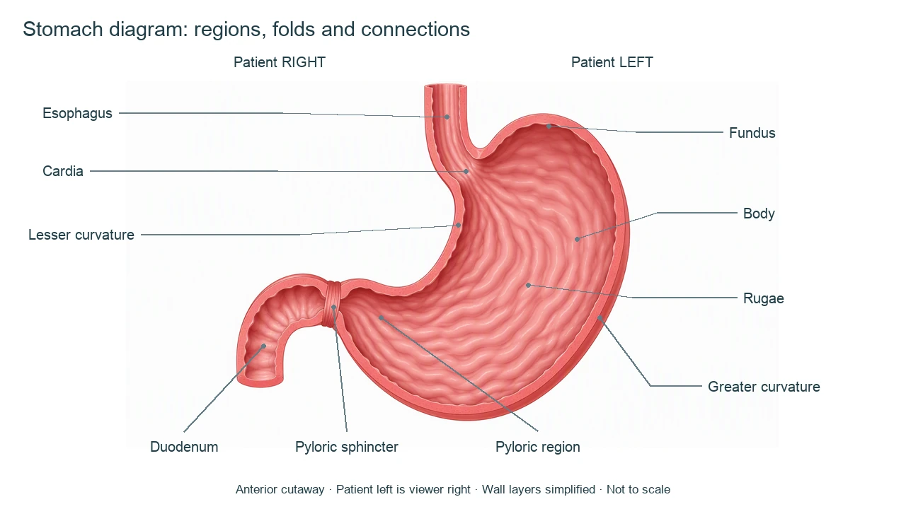
Stomach diagram labels and anatomical landmarks
Use this label key to plan your diagram. Soft tissues or functions described in the table may require an additional view or layer.
| Label | Where to find it | Role or drawing check |
|---|---|---|
| Esophagus and cardia | Inlet at the gastroesophageal junction | The esophagus delivers a swallowed bolus; the cardia is the stomach region around its opening. |
| Fundus | Dome above the cardia on patient left | A superior region, not the pyloric outlet. In this anterior view it appears toward viewer right. |
| Body | Largest central region | Provides space for storage and mixing with gastric secretions. |
| Lesser curvature | Shorter concave border | Runs from the cardia toward the pyloric region; do not confuse it with the convex outer curve. |
| Greater curvature | Longer convex border | Forms the broad outer border around the fundus and body. |
| Rugae | Folds on the internal surface | Folds of mucosa and submucosa that flatten as the stomach distends; they are not villi. |
| Pyloric region and sphincter | Distal narrowing near the outlet | The antrum leads toward the pyloric canal and muscular sphincter, which regulates emptying. |
| Duodenum | First small-intestinal segment beyond the pylorus | Receives chyme from the stomach; it is not part of the stomach itself. |
Anatomical relationships: OpenStax: The Stomach. On small screens, scroll the table horizontally.
Read a stomach diagram beyond the outline
A stomach diagram can explain orientation, regions and food movement. Secretory cells and muscle layers need their own enlarged section.
01
Regions are not separate chambers
Cardia, fundus, body and pyloric region are continuous parts of one lumen, without partitions between every label.
02
Rugae help accommodate distension
The internal folds flatten as volume increases. Gastric pits and glands are microscopic structures and cannot be identified from a broad fold alone.
03
Mixing precedes regulated emptying
Muscular contractions mix contents into chyme. The pyloric outlet controls delivery to the duodenum; most nutrient absorption occurs downstream in the small intestine.
Common stomach diagram mistakes
Reversing the fundus and pylorus
In this anterior stomach diagram, the fundus lies toward viewer right and the pyloric outlet toward viewer left.
Labeling rugae as villi
Stomach rugae are broad folds; intestinal villi are different structures and should not be drawn as the same feature.
Adding a hard valve at every region
The cardia is a region. The lower esophageal sphincter is primarily a functional high-pressure zone, unlike an exaggerated ring at every named boundary.
Create your own stomach diagram
Start with the prefilled prompt in the generator above. Reading the reference is free; creating a new AI draft requires an account and available credits. See current pricing.
Choose Science Drawing for an illustrated draft, or Editable Diagram when you need separate shapes and text labels. Check the anatomy in either mode before using the result.
- 1
Choose the orientation
Request an anterior stomach diagram with patient left explicitly marked.
- 2
Build the main label list
Start with inlet, four regions, curvatures, rugae and duodenal outlet; add a separate wall inset if needed.
- 3
Review digestive context
Check the pyloric connection and keep organ-level labels separate from glands or cellular secretions.
A prompt you can adapt
Create a labeled stomach diagram in anterior cutaway with patient LEFT on the viewer’s right. Show one esophageal inlet into the cardia, a fundus dome above it toward patient left, the large body, a shorter concave lesser curvature, longer convex greater curvature, internal rugae, pyloric antrum and canal, a pyloric sphincter and short duodenal outlet toward patient right. Do not add intestinal villi or partitions between stomach regions. Keep gastric gland and wall-layer detail in a separate inset if requested. White background, clear noncrossing leaders, not to scale.
Use this prompt in the generatorStomach FAQ
What are the four regions of a stomach diagram?
The four main regions are cardia, fundus, body and pyloric region. The pyloric region includes the antrum and canal leading to the pylorus.
Where is the fundus in an anterior stomach diagram?
The fundus is the dome superior to the cardia on patient left, which appears toward the viewer’s right in this anterior view.
What are rugae?
Rugae are folds of the stomach mucosa and submucosa. They flatten as the stomach fills and are not intestinal villi.
What connects the stomach to the small intestine?
The pyloric outlet leads into the duodenum, the first part of the small intestine. The pyloric sphincter regulates gastric emptying.
Does the stomach absorb most nutrients?
No. The stomach stores and mixes contents and begins important digestive processes, while most nutrient absorption occurs in the small intestine.
How is this different from a digestive system diagram?
This stomach diagram focuses on one organ and its landmarks. A digestive system diagram shows the broader alimentary route and accessory organs.
Can I generate an editable stomach diagram?
Yes. Choose Editable Diagram for separate shapes and labels, or Science Drawing for an illustrated draft. Review orientation and connections before use. Generation requires an account and credits.
Sources and editorial notes
- OpenStax, Anatomy and Physiology: The Stomach — factual background for the accompanying explanation.
- OpenStax: Digestive System Processes and Regulation — additional educational context.
Prepared by the AcaDraw product team. Source and label check: . Independent medical review has not been completed. This material is for education; AI drafts need review against reliable anatomy references before use and are not for diagnosis or treatment.
Turn the reference into your own teaching figure
Choose the structures your lesson needs, create a draft, and review it before sharing.
Read the stomach regions and rugae guideKidney diagram
Explore the labeled reference and drawing guide.
Nephron diagram
Explore the labeled reference and drawing guide.
Female reproductive system diagram
Explore the labeled reference and drawing guide.
Lungs diagram
Explore the labeled reference and drawing guide.
Nervous system diagram
Explore the labeled reference and drawing guide.
Labeled heart diagram
Explore the labeled reference and drawing guide.
Foot Anatomy diagram
Explore the labeled reference and drawing guide.
Shoulder Anatomy diagram
Explore the labeled reference and drawing guide.
Labeled brain diagram
Explore the labeled reference and drawing guide.
Digestive system diagram
Explore the labeled reference and drawing guide.
Respiratory system diagram
Explore the labeled reference and drawing guide.
Skeletal system diagram
Explore the labeled reference and drawing guide.
Neuron diagram
Explore the labeled reference and drawing guide.
Medical illustration generator
Plan other educational anatomy illustrations.