Ear Anatomy Diagram Generator
Create an ear anatomy diagram that follows sound from the outer ear through the middle ear to the cochlea and auditory nerve. Keep balance structures visible without confusing them with the hearing pathway.
SEE ACADRAW IN ACTION
Science drawing, from idea to editable figure
Create a science illustration with AI. Refine drawings, build editable diagrams, and convert images to SVG.
Demos play as you scroll into view. Choose a feature to explore.
Trusted by students & researchers from 480+ universities
A global community spanning 60+ countries and regions, from QS top-ranked universities to leading research institutes
























University logos belong to their respective institutions and are shown solely to illustrate our user community. They do not imply any partnership or endorsement.
Tool overview
Explore the use cases, workflow, and key things to know.
Follow sound first
Trace outer ear, canal, eardrum, ossicles, cochlea and auditory nerve in order.
Separate balance structures
Semicircular canals belong to the vestibular system; they are not the sound pathway.
Keep clinical claims out
A labeled anatomy figure does not measure hearing or diagnose loss.
Labeled reference
Ear anatomy diagram: outer, middle and inner ear
This ear anatomy diagram is a simplified side cutaway. It separates the sound pathway from the semicircular canals and does not represent hearing thresholds or a clinical finding.
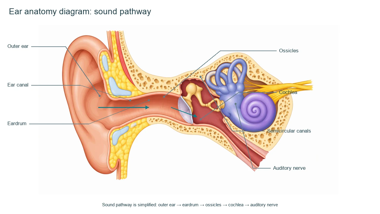
Ear Anatomy label key
Use this label key to plan your diagram. Soft tissues or functions described in the table may require an additional view or layer.
| Label or stage | Position or relationship | Meaning and drawing check |
|---|---|---|
| Outer ear | Pinna and entrance region | The outer ear collects and directs sound toward the canal. |
| Ear canal | Passage to the eardrum | The canal carries sound waves inward and protects the deeper structures. |
| Eardrum | Boundary between outer and middle ear | The tympanic membrane vibrates in response to sound. |
| Ossicles | Middle ear bones | Malleus, incus and stapes transmit and amplify vibration toward the inner ear. |
| Cochlea | Spiral inner-ear organ | The cochlea converts mechanical vibration into neural signals in the simplified model. |
| Semicircular canals | Inner-ear balance structures | They sense rotational movement and are shown separately from hearing. |
| Auditory nerve | Path toward the brain | The nerve carries auditory information away from the cochlea. |
Anatomical relationships: OpenStax: Ear anatomy. On small screens, scroll the table horizontally.
Read the ear anatomy diagram as two connected systems
Start with the hearing route, then identify the vestibular structures as a separate balance-related group.
01
Vibration and signals differ
The eardrum and ossicles transmit vibration; the cochlea and nerve support neural signaling.
02
Hearing and balance share the inner ear
The cochlea and semicircular canals have different roles despite being adjacent.
03
A side cutaway is selective
The diagram omits fine tissue layers and pressure details for readability.
Common ear anatomy mistakes
Calling the cochlea a bone
The cochlea is a spiral inner-ear organ, not one of the ossicles.
Putting the auditory nerve before the cochlea
In the simplified route, signals leave after the cochlea.
Using it to assess hearing
A diagram cannot replace audiometry or a clinical examination.
Create your own ear anatomy diagram
Start with the prefilled prompt in the generator above. Reading the reference is free; creating a new AI draft requires an account and available credits. See current pricing.
Choose Science Drawing for an illustrated draft, or Editable Diagram when you need separate shapes and text labels. Check the anatomy in either mode before using the result.
- 1
Choose the cutaway
Use a side section so outer, middle and inner ear structures fit on one canvas.
- 2
Trace the sound route
Place labels in the order sound or vibration travels.
- 3
Separate balance
Use a distinct note or color for semicircular canals and vestibular function.
A prompt you can adapt
Create a labeled ear anatomy diagram in side cutaway. Show outer ear, ear canal, eardrum, malleus/incus/stapes as ossicles, cochlea, semicircular canals and auditory nerve. Add arrows for the simplified sound pathway, keep labels editable and state that the figure is not for diagnosis.
Use this prompt in the generatorEar Anatomy FAQ
What are the main parts of an ear diagram?
A basic model includes outer ear, canal, eardrum, ossicles, cochlea and auditory nerve, with semicircular canals for context.
What do the ossicles do?
They transmit and amplify vibration from the eardrum toward the inner ear.
Are semicircular canals part of hearing?
They are primarily associated with balance and rotational movement, not the main sound pathway.
Is this a clinical ear exam?
No. It is a simplified educational diagram and cannot diagnose hearing or balance problems.
Sources and editorial notes
- OpenStax, Anatomy and Physiology: Ear anatomy — factual background for the accompanying explanation.
- OpenStax: The Ear — additional educational context.
Prepared by the AcaDraw product team. Source and label check: . Independent medical review has not been completed. This material is for education; AI drafts need review against reliable anatomy references before use and are not for diagnosis or treatment.
Turn the reference into your own teaching figure
Choose the structures your lesson needs, create a draft, and review it before sharing.
Read the ear anatomy guideSkin Diagram
Explore the labeled reference and drawing guide.
Urinary System Diagram
Explore the labeled reference and drawing guide.
Endocrine System Diagram
Explore the labeled reference and drawing guide.
Eye Anatomy Diagram
Explore the labeled reference and drawing guide.
Human Anatomy Diagram
Explore the labeled reference and drawing guide.
Lymphatic System Diagram
Explore the labeled reference and drawing guide.
Muscular System Diagram
Explore the labeled reference and drawing guide.
Kidney diagram
Explore the labeled reference and drawing guide.
Nephron diagram
Explore the labeled reference and drawing guide.
Stomach diagram
Explore the labeled reference and drawing guide.
Female reproductive system diagram
Explore the labeled reference and drawing guide.
Lungs diagram
Explore the labeled reference and drawing guide.
Nervous system diagram
Explore the labeled reference and drawing guide.
Labeled heart diagram
Explore the labeled reference and drawing guide.
Foot Anatomy diagram
Explore the labeled reference and drawing guide.
Shoulder Anatomy diagram
Explore the labeled reference and drawing guide.
Labeled brain diagram
Explore the labeled reference and drawing guide.
Digestive system diagram
Explore the labeled reference and drawing guide.
Respiratory system diagram
Explore the labeled reference and drawing guide.
Skeletal system diagram
Explore the labeled reference and drawing guide.
Neuron diagram
Explore the labeled reference and drawing guide.
Medical illustration generator
Plan other educational anatomy illustrations.