Brainstem Diagram Generator
Explore a labeled brainstem diagram showing the midbrain, pons and medulla, then create a focused educational draft with clear boundaries. Use a focused scope, review labels and connections, and keep the reference qualitative.
SEE ACADRAW IN ACTION
Science drawing, from idea to editable figure
Create a science illustration with AI. Refine drawings, build editable diagrams, and convert images to SVG.
Demos play as you scroll into view. Choose a feature to explore.
Trusted by students & researchers from 480+ universities
A global community spanning 60+ countries and regions, from QS top-ranked universities to leading research institutes























University logos belong to their respective institutions and are shown solely to illustrate our user community. They do not imply any partnership or endorsement.
Tool overview
Explore the use cases, workflow, and key things to know.
Define the scope
Choose the view, audience and labels before generating.
Keep relationships explicit
Use readable arrows, connectors or boundaries instead of decorative ambiguity.
Review before sharing
Check spelling, endpoints and claims against reliable references.
Labeled reference
Brainstem reference: clear labels and relationships
Explore a labeled brainstem diagram showing the midbrain, pons and medulla, then create a focused educational draft with clear boundaries. The original teaching illustration is schematic and does not encode measured data or guarantee factual correctness.
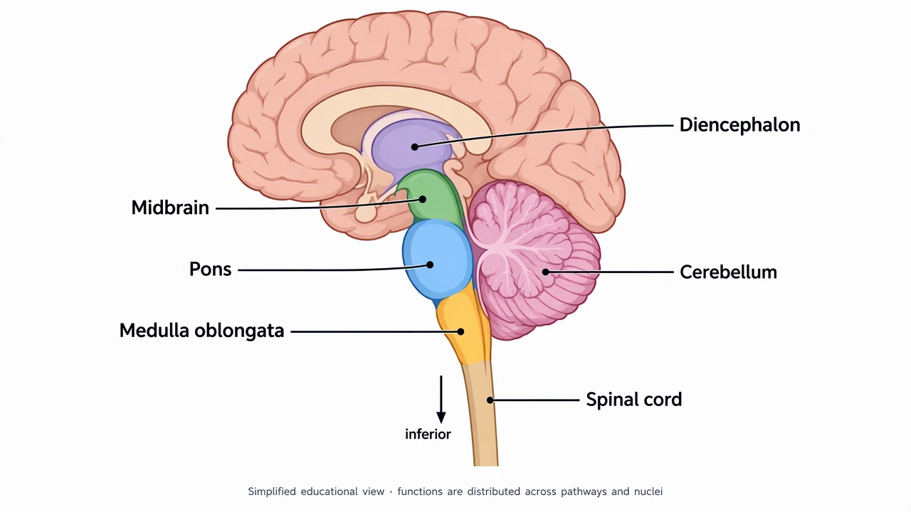
Brain diagram labels: parts, locations and functions
Use this label key to plan your diagram. Soft tissues or functions described in the table may require an additional view or layer.
| Structure | Where to find it | Role and diagram note |
|---|---|---|
| Cerebrum and cerebral cortex | Large upper part of the brain; cortex is its outer gray-matter layer | Supports perception, voluntary action and higher cognitive functions through interconnected networks. The cortex is not the whole brain. |
| Frontal lobe | Anterior to the central sulcus | Includes regions involved in voluntary movement, planning and aspects of language. Functions are distributed across networks. |
| Parietal lobe | Behind the central sulcus and above the lateral sulcus | Includes the primary somatosensory cortex and supports sensory integration and spatial processing. |
| Temporal lobe | Below the lateral sulcus | Includes auditory processing regions; medial temporal structures contribute to memory. Those internal structures are not shown in the surface plate. |
| Occipital lobe | Posterior end of the cerebrum | Contains primary visual cortex and other visual processing regions. Do not place it next to the forehead. |
| Central sulcus | Groove between frontal and parietal lobes | Separates the precentral gyrus, which contains primary motor cortex, from the postcentral gyrus, which contains primary somatosensory cortex. |
| Cerebellum | Behind the brainstem, below the posterior cerebrum | Contributes to coordination, balance and motor learning; visible as the smaller folded structure in the lateral plate. It is not a cerebral lobe. |
| Brainstem | Between the diencephalon and spinal cord | Comprises midbrain, pons and medulla. The second reference labels these regions; the spinal cord is not part of the brainstem. |
| Thalamus and hypothalamus | Deep structures of the diencephalon, above the brainstem | The thalamus participates in relay and processing; the hypothalamus contributes to homeostasis and endocrine regulation. Add a separate labeled internal view to show them. |
Anatomical relationships: OpenStax: The central nervous system. On small screens, scroll the table horizontally.
Choose the right view for your brain diagram
A surface drawing, a medial section and a frontal view reveal different relationships. Do not put internal labels on an intact outer surface without explaining a cutaway.
01
Scope controls meaning
A focused view is easier to read when the audience and intended lesson are explicit.
02
Labels need endpoints
Place each label next to the structure, role or relationship it describes.
03
Qualitative by default
This reference does not calculate or display a real dataset.
Common brain diagram mistakes
Overloading the page
Keep secondary details in an inset or a separate figure.
Leaving relationships implicit
Label lines and boundaries so readers can follow the intended meaning.
Treating a draft as verified
Review AI-generated text and structure before publishing.
Create your own brainstem diagram
Start with the prefilled prompt in the generator above. Reading the reference is free; creating a new AI draft requires an account and available credits. See current pricing.
Choose Science Drawing for an illustrated draft, or Editable Diagram when you need separate shapes and text labels. Check the anatomy in either mode before using the result.
- 1
Choose scope and view
Start with the four lobes for an introductory worksheet. Request a separate section when you need deeper structures.
- 2
Generate clear label groups
Keep surface regions, internal structures and brainstem parts in distinct panels. Use a short legend rather than dense functional claims.
- 3
Check anatomy and export
Verify orientation, sulci and every leader-line endpoint. Correct the draft against reliable references before sharing or exporting.
A prompt you can adapt
Create a clean, editable educational brainstem with short readable labels, generous spacing and explicit relationships. Keep the model qualitative, avoid invented measurements, and add a small scope note.
Use this prompt in the generatorBrainstem FAQ
What are the main parts of the brain?
A useful adult-brain overview distinguishes the cerebrum, diencephalon, brainstem and cerebellum. The cerebral cortex is the outer gray-matter layer of the cerebrum; it is not a synonym for the entire brain.
What are the four lobes in a lateral brain diagram?
The frontal, parietal, temporal and occipital lobes are the four lobes commonly shown on the outer lateral surface. The insula is hidden deep to the lateral sulcus, so a complete anatomical classification needs more detail than four colored surface regions.
What is the difference between the cerebrum and cerebellum?
The cerebrum forms the large paired cerebral hemispheres. The cerebellum is a separate structure behind the brainstem that contributes to coordination, balance and motor learning.
Can I see the thalamus in the outer brain view?
Not on the intact lateral surface. Use an appropriate section or cutaway to show the thalamus and other deep structures, and state which view the diagram uses.
How do I make a blank brain diagram for labeling practice?
Create and check your own labeled draft, duplicate it, and remove the names from the copy. Keep numbered pointers and a separate answer key.
Is the brain diagram generator free?
The labeled reference is free to read. Creating a new AI draft requires signing in and available credits. Choose Science Drawing for an illustrated draft or Editable Diagram for separate shapes and labels, then review the anatomy before use.
Sources and editorial notes
- OpenStax, Anatomy and Physiology: The central nervous system — factual background for the accompanying explanation.
- NINDS: Brain Basics — Know Your Brain — additional educational context.
Prepared by the AcaDraw product team. Source and label check: . Independent medical review has not been completed. This material is for education; AI drafts need review against reliable anatomy references before use and are not for diagnosis or treatment.
Turn the reference into your own teaching figure
Choose the structures your lesson needs, create a draft, and review it before sharing.
Read the step-by-step tutorialSkin Diagram
Explore the labeled reference and drawing guide.
Urinary System Diagram
Explore the labeled reference and drawing guide.
Endocrine System Diagram
Explore the labeled reference and drawing guide.
Eye Anatomy Diagram
Explore the labeled reference and drawing guide.
Human Anatomy Diagram
Explore the labeled reference and drawing guide.
Ear Anatomy Diagram
Explore the labeled reference and drawing guide.
Lymphatic System Diagram
Explore the labeled reference and drawing guide.
Muscular System Diagram
Explore the labeled reference and drawing guide.
Kidney diagram
Explore the labeled reference and drawing guide.
Nephron diagram
Explore the labeled reference and drawing guide.
Stomach diagram
Explore the labeled reference and drawing guide.
Female reproductive system diagram
Explore the labeled reference and drawing guide.
Lungs diagram
Explore the labeled reference and drawing guide.
Nervous system diagram
Explore the labeled reference and drawing guide.
Labeled heart diagram
Explore the labeled reference and drawing guide.
Foot Anatomy diagram
Explore the labeled reference and drawing guide.
Shoulder Anatomy diagram
Explore the labeled reference and drawing guide.
Labeled brain diagram
Explore the labeled reference and drawing guide.
Digestive system diagram
Explore the labeled reference and drawing guide.
Respiratory system diagram
Explore the labeled reference and drawing guide.
Skeletal system diagram
Explore the labeled reference and drawing guide.
Neuron diagram
Explore the labeled reference and drawing guide.
Medical illustration generator
Plan other educational anatomy illustrations.