Nervous System Diagram Generator
Create a nervous system diagram that separates the central and peripheral divisions. Locate brain, spinal cord and peripheral nerves, then use a clear legend to explain sensory input and motor output.
SEE ACADRAW IN ACTION
Science drawing, from idea to editable figure
Create a science illustration with AI. Refine drawings, build editable diagrams, and convert images to SVG.
Demos play as you scroll into view. Choose a feature to explore.
Trusted by students & researchers from 480+ universities
A global community spanning 60+ countries and regions, from QS top-ranked universities to leading research institutes























University logos belong to their respective institutions and are shown solely to illustrate our user community. They do not imply any partnership or endorsement.
Tool overview
Explore the use cases, workflow, and key things to know.
Read the nervous system diagram by division
Identify the brain and spinal cord as CNS, with peripheral nerves and ganglia as PNS.
Separate anatomy from function
CNS/PNS describes location; sensory/motor describes information flow relative to the CNS.
Connect scales without mixing them
Use this body-level map alongside a brain reference and a separate neuron diagram.
Labeled reference
Nervous system diagram: central structures and peripheral branches
This nervous system diagram is an orientation map, not a complete nerve atlas. Violet marks the brain and spinal cord; teal marks schematic peripheral branches. Many nerves, ganglia and pathways are omitted. The spinal cord stops in the upper lumbar region in the typical adult, while peripheral nerve roots continue below; body proportions and branch positions are simplified.
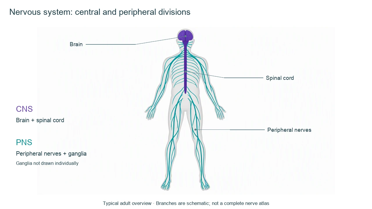
Nervous system diagram label key
Use this label key to plan your diagram. Soft tissues or functions described in the table may require an additional view or layer.
| Label | Anatomical division or position | Function and reading note |
|---|---|---|
| Brain | CNS; inside the skull | Integrates information and coordinates many functions. Its major regions need a separate enlarged diagram. |
| Spinal cord | CNS; within the vertebral canal | Carries pathways and contains circuits for spinal reflexes. In the typical adult it ends around vertebral levels L1–L2. |
| Peripheral nerves | PNS; outside the brain and spinal cord | Bundles of axons connecting the CNS with peripheral structures. They are not blood vessels. |
| Cranial nerves | Usually discussed with the PNS | Twelve pairs are conventionally named. The optic nerve has CNS characteristics, so a broad teaching classification has exceptions. |
| Spinal nerves | PNS; connected to spinal roots | There are 31 pairs. A spinal nerve is generally mixed after sensory and motor roots join. This map does not show every pair. |
| Ganglia | PNS; clusters of neuron cell bodies | Distinguish a ganglion from a nerve bundle. Individual ganglia are omitted in the overview. |
| Sensory / afferent pathways | Directed toward the CNS | Carry information from receptors; this is a functional direction, not a third anatomical division. |
| Motor / efferent pathways | Directed away from the CNS | Carry output toward effectors. Somatic motor output supplies skeletal muscle; autonomic output regulates smooth muscle, cardiac muscle and glands. |
Anatomical relationships: OpenStax: Basic Structure and Function of the Nervous System. On small screens, scroll the table horizontally.
Follow a pathway through a nervous system diagram
Use a nervous system diagram to ask where a structure belongs and which way information travels. Those are separate questions and need separate labels.
01
Anatomical map: CNS and PNS
The brain and spinal cord form the CNS. Peripheral nerves and ganglia belong to the PNS. A body overview cannot show every named nerve, so its legend should identify the level of simplification.
02
Functional map: input and output
Afferent means toward the CNS and efferent means away from it. Many peripheral nerves contain both sensory and motor axons; a single color does not mean all signals in that nerve travel one way.
03
Motor organization: somatic and autonomic
Somatic motor pathways control skeletal muscle. Autonomic pathways regulate cardiac muscle, smooth muscle and glands; sympathetic and parasympathetic divisions are part of that discussion. Enteric circuits need explicit context rather than a misleading third branch beside the entire CNS and PNS.
Nervous system diagram mistakes to check
Extending the cord to the tailbone
In a typical adult the spinal cord ends around L1–L2 vertebral levels. Nerve roots continue below it; do not label those roots as spinal cord.
Equating PNS with autonomic
The PNS includes sensory pathways and somatic motor connections as well as autonomic components.
Drawing every nerve as one-way
Mixed peripheral nerves carry different axons in different directions. Use separately labeled afferent and efferent arrows in a pathway inset.
Create your own nervous system diagram
Start with the prefilled prompt in the generator above. Reading the reference is free; creating a new AI draft requires an account and available credits. See current pricing.
Choose Science Drawing for an illustrated draft, or Editable Diagram when you need separate shapes and text labels. Check the anatomy in either mode before using the result.
- 1
Choose a body map or pathway
Use a nervous system diagram overview for CNS/PNS locations; request a separate flow panel for sensory and motor arrows.
- 2
Define the legend
Specify distinct CNS and PNS colors, a typical adult body model and the labels you need.
- 3
Review boundaries and direction
Check cord termination, nerve labels and the meaning of afferent/efferent before publishing the draft.
A prompt you can adapt
Create an educational nervous system diagram in a simplified adult anterior body outline. Use violet for brain and spinal cord (CNS) and teal for schematic peripheral nerves (PNS). End the spinal cord in the upper lumbar region, not the pelvis; show peripheral roots and branches continuing below without labeling them spinal cord. Label brain, spinal cord, peripheral nerves and a clear CNS/PNS legend. State that not all nerves or ganglia are shown. Add a separate small pathway panel only if needed: receptor → sensory/afferent pathway → CNS → motor/efferent pathway → effector. Do not imply every peripheral nerve carries signals in only one direction. No clinical diagnosis.
Use this prompt in the generatorNervous system FAQ
What are the two main divisions in a nervous system diagram?
The central nervous system (CNS) comprises brain and spinal cord. The peripheral nervous system (PNS) includes peripheral nerves and ganglia. This is an anatomical classification.
What should a basic nervous system diagram label?
Label the brain, spinal cord and peripheral nerves, with a legend separating CNS and PNS. Use separate insets for ganglia, detailed cranial nerves or functional pathways.
Are afferent and efferent the same as CNS and PNS?
No. Afferent and efferent describe direction relative to the CNS: toward it or away from it. CNS and PNS describe anatomical divisions.
Does the spinal cord extend the full length of the back?
Not in the typical adult. It ends around L1–L2 vertebral levels, while lower spinal nerve roots continue within the canal as the cauda equina.
Is the autonomic nervous system all of the PNS?
No. Autonomic pathways are only part of nervous system organization. The PNS also includes sensory pathways and somatic motor connections. Autonomic control also involves central structures.
How is this different from a neuron diagram?
A nervous system diagram is a body-level organization map. A neuron diagram shows the structure of one cell, such as soma, dendrites and axon.
Can I make an editable nervous system diagram?
Yes. Choose Editable Diagram for shapes and labels or Science Drawing for an illustrated draft. Generation requires an account and credits. Review the anatomy and arrow directions against reliable references.
Sources and editorial notes
- OpenStax, Anatomy and Physiology: Basic Structure and Function of the Nervous System — factual background for the accompanying explanation.
- OpenStax: The Function of Nervous Tissue — additional educational context.
Prepared by the AcaDraw product team. Source and label check: . Independent medical review has not been completed. This material is for education; AI drafts need review against reliable anatomy references before use and are not for diagnosis or treatment.
Turn the reference into your own teaching figure
Choose the structures your lesson needs, create a draft, and review it before sharing.
Read the CNS, PNS and pathways guideFemale reproductive system diagram
Explore the labeled reference and drawing guide.
Lungs diagram
Explore the labeled reference and drawing guide.
Labeled heart diagram
Explore the labeled reference and drawing guide.
Foot Anatomy diagram
Explore the labeled reference and drawing guide.
Shoulder Anatomy diagram
Explore the labeled reference and drawing guide.
Labeled brain diagram
Explore the labeled reference and drawing guide.
Digestive system diagram
Explore the labeled reference and drawing guide.
Respiratory system diagram
Explore the labeled reference and drawing guide.
Skeletal system diagram
Explore the labeled reference and drawing guide.
Neuron diagram
Explore the labeled reference and drawing guide.
Medical illustration generator
Plan other educational anatomy illustrations.