Kidney Diagram Generator
Create a kidney diagram that separates the cortex, medullary pyramids and urine-collecting spaces. Explore a labeled cutaway, follow drainage toward the ureter and build your own anatomy teaching draft.
SEE ACADRAW IN ACTION
Science drawing, from idea to editable figure
Create a science illustration with AI. Refine drawings, build editable diagrams, and convert images to SVG.
Demos play as you scroll into view. Choose a feature to explore.
Trusted by students & researchers from 480+ universities
A global community spanning 60+ countries and regions, from QS top-ranked universities to leading research institutes
























University logos belong to their respective institutions and are shown solely to illustrate our user community. They do not imply any partnership or endorsement.
Tool overview
Explore the use cases, workflow, and key things to know.
Read the kidney diagram in layers
Identify the capsule, cortex and medulla before naming the central collecting spaces.
Trace the drainage route
Follow calyces into the renal pelvis and ureter; keep this route separate from blood flow.
Switch scale for filtration
Use a nephron diagram when the lesson needs the glomerulus, tubules or reabsorption.
Labeled reference
Labeled kidney diagram: from cortex to renal pelvis
This kidney diagram is a simplified longitudinal cutaway. The outer cortex surrounds the darker medullary pyramids. Collecting cups receive urine at the pyramid tips and converge toward the renal pelvis and ureter. The illustration omits the renal vessels and microscopic nephrons so the tissue regions and drainage spaces remain distinct; it does not identify a patient side.
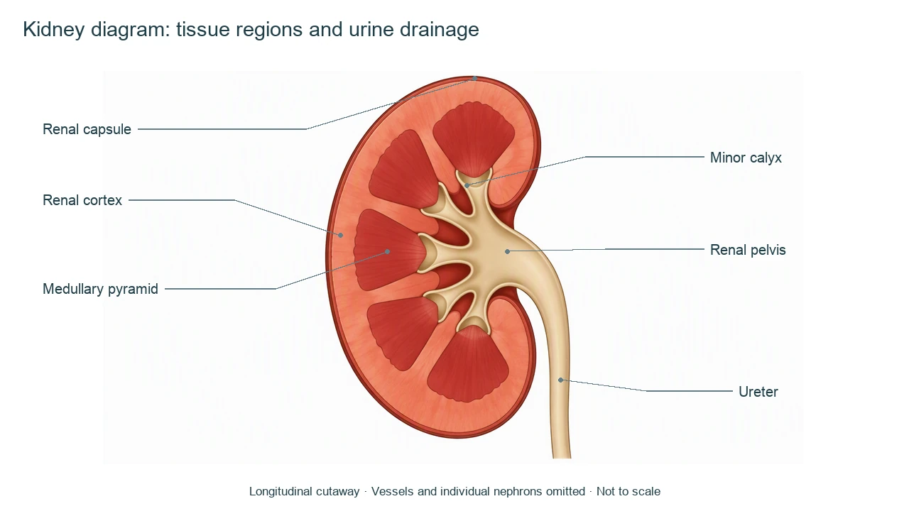
Kidney diagram labels: tissue regions and collecting spaces
Use this label key to plan your diagram. Soft tissues or functions described in the table may require an additional view or layer.
| Label | Where to find it | Function or drawing check |
|---|---|---|
| Renal capsule | Thin external covering | Fibrous covering distinct from the underlying cortex; the drawing exaggerates its thickness for visibility. |
| Renal cortex | Outer tissue below the capsule | Contains renal corpuscles and convoluted tubules; it is tissue, not a collecting chamber. |
| Renal medulla | Inner tissue organized into pyramids | Contains loops and collecting ducts. Its triangular regions are not empty sacs. |
| Medullary pyramid | Triangular medullary region | Its apex, the renal papilla, projects toward a minor calyx. The number drawn is illustrative. |
| Minor and major calyces | Collecting spaces near pyramid tips and centrally | Minor calyces receive urine from papillae; they join into major calyces leading toward the pelvis. |
| Renal pelvis | Central funnel near the medial hilum | Collects urine from the calyces and narrows into the ureter. |
| Ureter | Tube leaving the renal pelvis | Carries urine toward the bladder; it is not the urethra or a blood vessel. |
Anatomical relationships: OpenStax: Gross Anatomy of the Kidney. On small screens, scroll the table horizontally.
Read a kidney diagram at the organ level
A kidney diagram shows the location of tissues and collecting spaces. Filtration and transport need a microscopic view rather than extra arrows placed over the whole organ.
01
Cortex and medulla are tissues
Renal columns extend between pyramids. A pale central collecting region does not mean the entire medulla is hollow.
02
The drainage route starts after the tubules
Urine passes from papillary ducts into minor calyces, major calyces, the pelvis and the ureter. Filtration itself begins in renal corpuscles.
03
Blood flow needs a separate layer
Renal arteries supply blood and renal veins return it. This kidney diagram omits vessels, so a drainage arrow must not be read as the blood route.
Common kidney diagram mistakes
Using ureter and urethra interchangeably
The ureter runs from kidney to bladder; the urethra carries urine from the bladder outside the body.
Calling the pelvis the filtration site
The renal pelvis collects urine. Filtration across the glomerular barrier occurs at microscopic renal corpuscles.
Drawing pyramids as empty cups
Pyramids are medullary tissue; calyces are the collecting cups adjacent to their tips.
Create your own kidney diagram
Start with the prefilled prompt in the generator above. Reading the reference is free; creating a new AI draft requires an account and available credits. See current pricing.
Choose Science Drawing for an illustrated draft, or Editable Diagram when you need separate shapes and text labels. Check the anatomy in either mode before using the result.
- 1
Choose a cutaway view
Request a longitudinal kidney diagram and state whether vessels should be included.
- 2
Keep the labels at one scale
Use capsule, cortex, medulla, calyces, pelvis and ureter; place nephrons in a distinct inset.
- 3
Review the connections
Check that collecting cups join the pelvis and that the ureter continues from its funnel.
A prompt you can adapt
Create a labeled kidney diagram in longitudinal cutaway. Show a thin renal capsule, outer cortex, solid medullary pyramids with papillae, minor and major calyces, a central renal pelvis and one ureter. Keep cortical tissue distinct from collecting spaces. Use a separate inset if showing a nephron. Omit renal vessels for this drainage-focused overview, and do not show blood entering the pelvis. Clear noncrossing leaders, white background, simplified and not to scale.
Use this prompt in the generatorKidney FAQ
What should a labeled kidney diagram include?
An introductory kidney diagram labels the capsule, cortex, medulla or pyramids, calyces, renal pelvis and ureter. Vessels and a nephron inset can be added as separate layers.
What is the difference between cortex and medulla?
The cortex is the outer region containing renal corpuscles and convoluted tubules. The medulla is the inner region with pyramids, loops and collecting ducts.
Where does urine go after the collecting ducts?
Papillary ducts drain into minor calyces, which join major calyces, then the renal pelvis and ureter.
Where does filtration happen?
Filtration begins across the glomerular filtration barrier into the capsular space of renal corpuscles. It does not occur in the renal pelvis.
Is a kidney diagram the same as a nephron diagram?
No. A kidney diagram shows the organ and its regions. A nephron diagram enlarges the microscopic filtration and tubular pathway.
Can I make an editable kidney diagram?
Yes. Choose Editable Diagram for separate shapes and labels, or Science Drawing for an illustrated draft. Generation requires an account and credits; review the anatomy before use.
Sources and editorial notes
- OpenStax, Anatomy and Physiology: Gross Anatomy of the Kidney — factual background for the accompanying explanation.
- OpenStax: Microscopic Anatomy of the Kidney — additional educational context.
Prepared by the AcaDraw product team. Source and label check: . Independent medical review has not been completed. This material is for education; AI drafts need review against reliable anatomy references before use and are not for diagnosis or treatment.
Turn the reference into your own teaching figure
Choose the structures your lesson needs, create a draft, and review it before sharing.
Read the kidney anatomy and drainage guideNephron diagram
Explore the labeled reference and drawing guide.
Stomach diagram
Explore the labeled reference and drawing guide.
Female reproductive system diagram
Explore the labeled reference and drawing guide.
Lungs diagram
Explore the labeled reference and drawing guide.
Nervous system diagram
Explore the labeled reference and drawing guide.
Labeled heart diagram
Explore the labeled reference and drawing guide.
Foot Anatomy diagram
Explore the labeled reference and drawing guide.
Shoulder Anatomy diagram
Explore the labeled reference and drawing guide.
Labeled brain diagram
Explore the labeled reference and drawing guide.
Digestive system diagram
Explore the labeled reference and drawing guide.
Respiratory system diagram
Explore the labeled reference and drawing guide.
Skeletal system diagram
Explore the labeled reference and drawing guide.
Neuron diagram
Explore the labeled reference and drawing guide.
Medical illustration generator
Plan other educational anatomy illustrations.