Respiratory System Diagram Generator
Follow this labeled respiratory system diagram from the nasal cavity to the lungs. Separate the air-conducting passages from gas exchange, then create a focused and reviewable teaching draft.
SEE ACADRAW IN ACTION
Science drawing, from idea to editable figure
Create a science illustration with AI. Refine drawings, build editable diagrams, and convert images to SVG.
Demos play as you scroll into view. Choose a feature to explore.
Trusted by students & researchers from 480+ universities
A global community spanning 60+ countries and regions, from QS top-ranked universities to leading research institutes























University logos belong to their respective institutions and are shown solely to illustrate our user community. They do not imply any partnership or endorsement.
Tool overview
Explore the use cases, workflow, and key things to know.
Trace the air route
Connect the pharynx, larynx, trachea and branching airways.
Separate scale and function
Use an alveolar inset for gas exchange instead of hiding it in a torso view.
Check your teaching draft
Verify right and left sides, branches and arrow directions before export.
Labeled reference
Labeled respiratory system diagram: trace the airway
The reference combines a head section with a torso overview. Anatomical right is on the viewer’s left in the torso. It labels the main airways, lungs and diaphragm; smaller bronchioles and alveoli require an enlarged inset. The diaphragm helps move air by changing thoracic volume; air does not pass through this muscle.
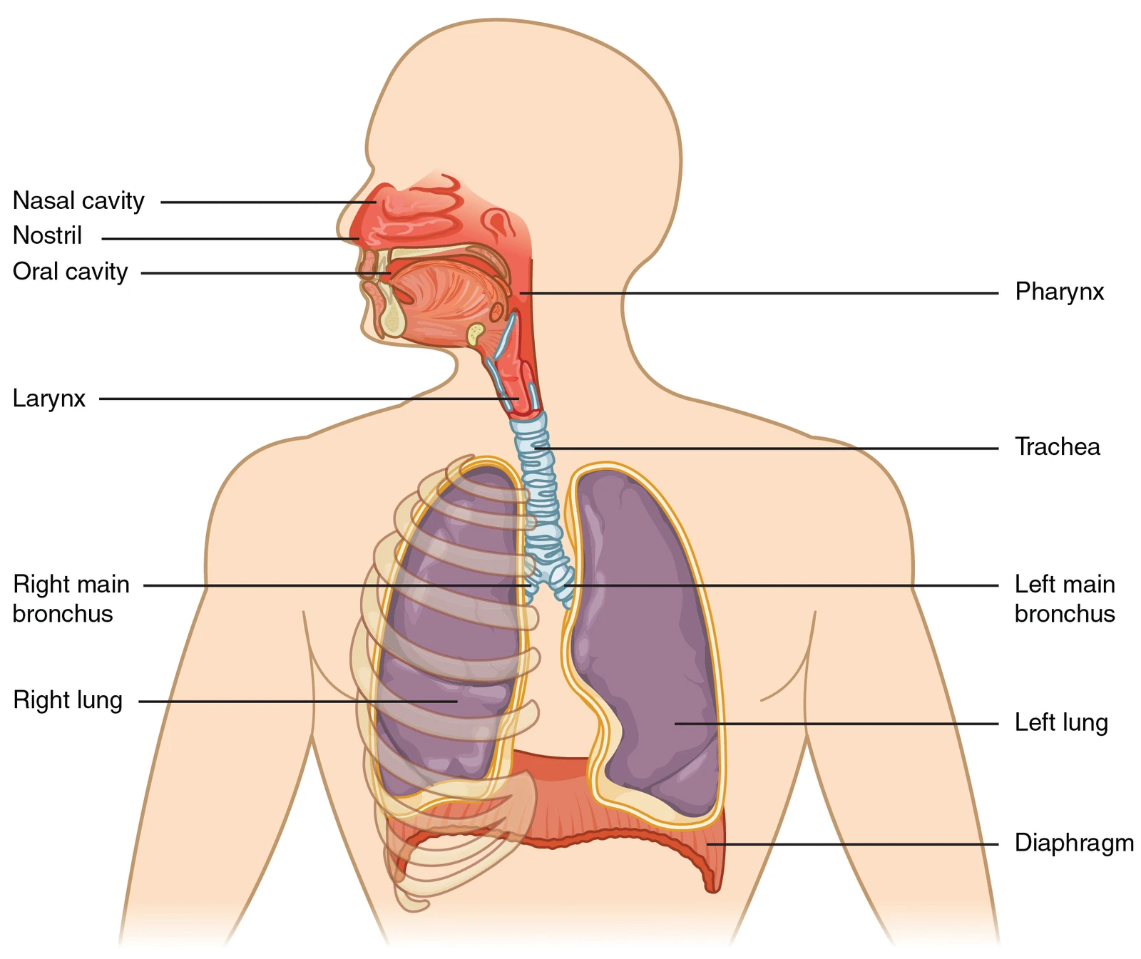
Respiratory system diagram labels and functions
Use this label key alongside the reference. Soft tissues or functions described in the table may require an additional view or layer.
| Structure | Position or connection | Function and drawing note |
|---|---|---|
| Nasal and oral cavities | Entrances that lead toward the pharynx | Air commonly enters through the nose, which helps filter, warm and humidify it. The mouth is another entrance. |
| Pharynx | Connects the nasal/oral region toward the larynx | Some pharyngeal regions are shared by food and air. Do not draw the airway as a continuation of the esophagus. |
| Larynx | Between the pharynx and trachea | Contains the vocal folds and helps protect the airway. It is distinct from the food passage behind it. |
| Trachea | Descends into the thorax before branching | Conducts air to the main bronchi. Cartilage helps maintain an open passage. |
| Right and left main bronchi | Branches entering the corresponding lungs | Continue dividing into smaller bronchi. Label sides relative to the person, not the viewer. |
| Bronchioles | Small airway branches within the lungs | Terminal bronchioles end the conducting zone; respiratory bronchioles begin the gas-exchanging region. Use an inset for these small branches. |
| Alveoli | Tiny air sacs associated with the respiratory zone | Oxygen diffuses from alveolar air toward pulmonary capillary blood; carbon dioxide moves the opposite way. Alveoli are not individually resolved in the overview. |
| Lungs and pleura | Right and left sides of the thorax | The right lung normally has three lobes and the left two. The pleura are membranes associated with the lungs and chest wall, not extra lung lobes. |
| Diaphragm | Muscular partition below the lungs | Contracts and descends during quiet inspiration, increasing thoracic volume. It is a breathing muscle, not an air conduit. |
Anatomical relationships: OpenStax: Organs and structures of the respiratory system. On small screens, scroll the table horizontally.
Read airflow and gas exchange in a respiratory system diagram
Use a respiratory system diagram at two scales: the body overview for air passage and a separate alveolar inset for diffusion. Keep airflow arrows distinct from arrows crossing the respiratory membrane.
01
Conducting zone
Nose, pharynx, larynx, trachea, bronchi and terminal bronchioles provide a route for air. They condition and distribute it without being the main gas-exchange surfaces.
02
Respiratory zone
Respiratory bronchioles, alveolar ducts and alveoli support gas exchange. An enlarged alveolar-capillary panel can show oxygen and carbon dioxide moving in opposite directions.
03
Ventilation and cellular respiration
Breathing moves air into and out of the lungs. Gas exchange moves gases between air and blood. Cellular respiration is a different set of metabolic processes in cells.
Common respiratory system diagram mistakes
Connecting the trachea to the stomach
Keep the trachea and esophagus distinct. The trachea branches into bronchi; the esophagus carries swallowed material toward the stomach.
Sending air through the diaphragm
Place the diaphragm beneath the lungs. Show its motion separately from arrows that follow the airway.
Reversing sides or gas arrows
Use anatomical right and left. In an alveolar inset, oxygen goes from air to blood and carbon dioxide from blood to air under normal pulmonary conditions.
Create your own respiratory system diagram
Start with the prefilled prompt in the generator above. Reading the reference is free; creating a new AI draft requires an account and available credits. See current pricing.
Choose Science Drawing for an illustrated draft, or Editable Diagram when you need separate shapes and text labels. Check the anatomy in either mode before using the result.
- 1
Choose the view and detail
Start with the main airway and lungs. Request a separate inset if your lesson needs bronchioles, alveoli or diffusion.
- 2
Specify different arrow meanings
Ask for airflow arrows inside passages and gas-transfer arrows across the alveolar-capillary barrier. Add a legend.
- 3
Review and make a labeling exercise
Check laterality, branching and diaphragm placement. Duplicate the corrected draft without names and retain an answer key.
A prompt you can adapt
Create a labeled educational human respiratory system diagram on a white background. Use a head section and anterior torso overview. Show nasal cavity, oral cavity, pharynx, larynx, trachea, right and left main bronchi, lungs and diaphragm. State anatomical right and left; show the right lung on the viewer’s left. Add a separate enlarged alveolar-capillary inset with oxygen moving from alveolar air to blood and carbon dioxide moving from blood to air. Keep airways separate from the esophagus. Do not route air through the diaphragm or connect it to the stomach. Indicate that the figure is simplified and not to scale. In Editable Diagram mode, keep shapes and labels separate for manual review.
Use this prompt in the generatorRespiratory System FAQ
What should a respiratory system diagram label?
A basic overview labels the nasal cavity, pharynx, larynx, trachea, main bronchi, lungs and diaphragm. Add an enlarged inset for bronchioles, alveoli and nearby capillaries when teaching gas exchange.
What is the order of the main air pathway?
Air travels through the nose or mouth, pharynx, larynx, trachea, bronchi and bronchioles toward the respiratory region and alveoli. Exhaled air returns along the passages in the opposite direction.
Where does gas exchange occur?
The respiratory zone includes respiratory bronchioles, alveolar ducts and alveoli. Across the alveolar-capillary barrier, oxygen normally moves toward blood and carbon dioxide toward alveolar air.
Does air pass through the diaphragm?
No. The diaphragm is a muscle below the lungs. Its contraction changes thoracic volume and helps produce the pressure difference that moves air.
Is breathing the same as cellular respiration?
No. Ventilation moves air, and gas exchange transfers gases. Cellular respiration refers to metabolic processes that release usable energy from nutrients.
Can I make an editable respiratory system diagram?
Yes. Use the prefilled prompt and choose Editable Diagram for separate shapes and labels. Generation needs an account and available credits. Review anatomy and gas-transfer arrows before using the draft.
Sources and editorial notes
- OpenStax, Anatomy and Physiology: Organs and structures of the respiratory system — primary anatomy reference and illustration source.
- NHLBI: How the Lungs Work — The Respiratory System — additional educational context.
Prepared by the AcaDraw product team. Source and label check: . Independent medical review has not been completed. This material is for education; AI drafts need review against reliable anatomy references before use and are not for diagnosis or treatment.
Turn the reference into your own teaching figure
Choose the structures your lesson needs, create a draft, and review it before sharing.
Read the step-by-step tutorialLabeled heart diagram
Explore the labeled reference and drawing guide.
Foot Anatomy diagram
Explore the labeled reference and drawing guide.
Shoulder Anatomy diagram
Explore the labeled reference and drawing guide.
Labeled brain diagram
Explore the labeled reference and drawing guide.
Digestive system diagram
Explore the labeled reference and drawing guide.
Medical illustration generator
Plan other educational anatomy illustrations.