Digestive System Diagram Generator
Follow this labeled digestive system diagram from mouth to anus. Separate the route taken by food from the accessory organs that supply secretions, then create a clear teaching draft.
SEE ACADRAW IN ACTION
Science drawing, from idea to editable figure
Create a science illustration with AI. Refine drawings, build editable diagrams, and convert images to SVG.
Demos play as you scroll into view. Choose a feature to explore.
Trusted by students & researchers from 480+ universities
A global community spanning 60+ countries and regions, from QS top-ranked universities to leading research institutes























University logos belong to their respective institutions and are shown solely to illustrate our user community. They do not imply any partnership or endorsement.
Tool overview
Explore the use cases, workflow, and key things to know.
Follow the food
Trace the mouth-to-anus pathway without detouring through accessory organs.
Know each organ’s role
Distinguish propulsion, digestion, nutrient absorption and elimination.
Review your own draft
Check organ positions, secretion arrows and label endpoints before export.
Labeled reference
Labeled digestive system diagram: trace the food pathway
This overview combines a head profile with an anterior torso view to expose the digestive organs. In the torso, the person’s right is on your left. Food travels through the alimentary canal; the liver, gallbladder and pancreas contribute secretions without being part of that route. The spleen is shown for orientation but is not a digestive organ.
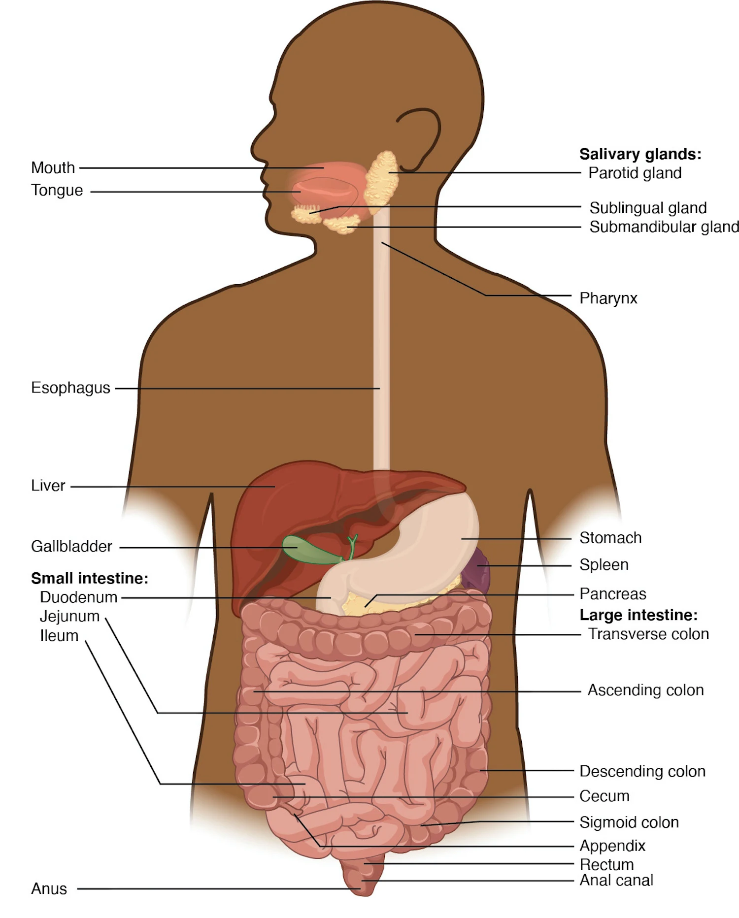
Digestive system diagram labels and organ functions
Use this label key alongside the reference. Soft tissues or functions described in the table may require an additional view or layer.
| Structure | Position or connection | Function and drawing note |
|---|---|---|
| Mouth and salivary glands | Beginning of the tract; glands drain into the mouth | Chewing mechanically breaks food down; saliva lubricates it and begins starch digestion. Food does not enter the salivary glands. |
| Pharynx and esophagus | Connect the mouth with the stomach | Swallowing and peristalsis move the bolus toward the stomach. Keep the esophagus distinct from the airway. |
| Stomach | Upper left abdomen of the person | Stores and mixes food; acid and enzymes begin substantial protein digestion. It empties into the duodenum, not the colon. |
| Small intestine | Duodenum → jejunum → ileum | Major site of chemical digestion and nutrient absorption. The ileum joins the large intestine at the ileocecal region. |
| Liver | Mainly in the person’s upper right abdomen | Produces bile, among many other functions. Food does not pass through the liver. |
| Gallbladder | Beneath the liver | Stores and concentrates bile, then releases it through ducts into the duodenum. It does not produce bile. |
| Pancreas | Mostly behind the stomach | Delivers digestive enzymes and bicarbonate through ducts to the duodenum. The overview exposes it; food does not pass through it. |
| Large intestine | Cecum → colon → rectum → anal canal | Absorbs remaining water and electrolytes and forms, stores and moves feces. Most nutrient absorption occurs earlier in the small intestine. |
| Rectum and anus | Terminal part of the digestive tract | The rectum stores feces before defecation; the anal canal leads to the anus. Do not connect the tract to the urinary bladder. |
| Spleen | Near the stomach in the reference | A lymphatic organ included as an anatomical neighbor, not a digestive gland or part of the food pathway. |
Anatomical relationships: OpenStax: Overview of the digestive system. On small screens, scroll the table horizontally.
Separate food movement from digestive secretions
In a digestive system diagram, use one arrow style for food moving along the tract and another for secretions entering it. This keeps the food pathway distinct from the inputs supplied by accessory organs.
01
Food through the tract
Mouth → pharynx → esophagus → stomach → duodenum → jejunum → ileum → cecum → ascending, transverse, descending and sigmoid colon → rectum → anal canal → anus.
02
Accessory organs into the tract
Salivary glands release saliva into the mouth. Bile from the liver, stored in the gallbladder, and pancreatic secretions reach the duodenum through ducts. These are input pathways, not the route of food.
03
Digestion and absorption
Digestion breaks food into smaller components. Absorption moves substances across the gut lining. Most nutrients are absorbed in the small intestine; the large intestine recovers remaining water and electrolytes and processes feces.
Common digestive system diagram mistakes
Sending food into the liver or pancreas
Connect these organs to the duodenum using secretion arrows. The food pathway stays inside the alimentary canal.
Mirroring the abdominal organs
In an anterior view, the liver is mainly on the viewer’s left and the stomach on the viewer’s right. State anatomical sides before placing labels.
Skipping the small intestine
The stomach empties into the duodenum. Show the small intestine connecting to the cecum; do not join the stomach directly to the large intestine.
Create your own digestive system diagram
Start with the prefilled prompt in the generator above. Reading the reference is free; creating a new AI draft requires an account and available credits. See current pricing.
Choose Science Drawing for an illustrated draft, or Editable Diagram when you need separate shapes and text labels. Check the anatomy in either mode before using the result.
- 1
Choose a teaching level
Use a short organ list for a first lesson, or add intestinal subdivisions and accessory-organ ducts for an advanced worksheet.
- 2
Draw two kinds of connection
Ask for a continuous food route plus distinct secretion inputs. Avoid combining every chemical process into the anatomy panel.
- 3
Review and prepare a worksheet
Check laterality, the stomach-to-duodenum connection, the ileocecal junction and duct endpoints. Duplicate a checked draft to make a blank labeling exercise.
A prompt you can adapt
Create a clear educational human digestive system diagram on a white background. Use a head profile with an anterior torso overview and label the view. Show mouth, pharynx, esophagus, stomach, duodenum, jejunum, ileum, cecum, ascending colon, transverse colon, descending colon, sigmoid colon, rectum and anus. Place the liver mainly on the person’s right and stomach on the person’s left. Add liver, gallbladder, pancreas and salivary glands as accessory organs. Use distinct arrows for the food route and for secretions entering the mouth or duodenum. Do not route food through accessory organs, the spleen or the urinary system. Use spaced labels and no disease findings. Keep shapes and labels separate in Editable Diagram mode for manual review.
Use this prompt in the generatorDigestive System FAQ
What is the correct order of the digestive system?
For the main food route: mouth, pharynx, esophagus, stomach, small intestine, large intestine, rectum, anal canal and anus. The small intestine is duodenum, jejunum and ileum. A detailed large-intestine route begins at the cecum and continues through the colon to the rectum.
Does food pass through the liver, gallbladder or pancreas?
No. These are accessory organs. They supply or store secretions that enter the digestive tract through ducts; food remains in the alimentary canal.
Where does most nutrient absorption happen?
Most nutrient absorption occurs in the small intestine. The large intestine absorbs remaining water and electrolytes and processes feces. Water absorption also occurs in the small intestine.
Is bile made in the gallbladder?
No. The liver makes bile. The gallbladder stores and concentrates it and releases it into the duodenum. Bile helps disperse fats; it is not a digestive enzyme.
Why is the spleen labeled in the reference?
It is a neighboring organ shown for anatomical orientation. The spleen belongs to the lymphatic system and is not part of the digestive tract or an accessory digestive gland.
Can I make an editable digestive system diagram?
Yes. Use the prefilled prompt above and choose Editable Diagram when you need separate shapes and text labels. Generation requires an account and available credits. Check the anatomy and all arrow endpoints against reliable references before using the result.
Sources and editorial notes
- OpenStax, Anatomy and Physiology: Overview of the digestive system — primary anatomy reference and illustration source.
- NIDDK: Your Digestive System & How It Works — additional educational context.
Prepared by the AcaDraw product team. Source and label check: . Independent medical review has not been completed. This material is for education; AI drafts need review against reliable anatomy references before use and are not for diagnosis or treatment.
Turn the reference into your own teaching figure
Choose the structures your lesson needs, create a draft, and review it before sharing.
Read the step-by-step tutorialLabeled heart diagram
Explore the labeled reference and drawing guide.
Foot Anatomy diagram
Explore the labeled reference and drawing guide.
Shoulder Anatomy diagram
Explore the labeled reference and drawing guide.
Labeled brain diagram
Explore the labeled reference and drawing guide.
Medical illustration generator
Plan other educational anatomy illustrations.