Urinary System Diagram Generator
Create a urinary system diagram that follows urine from the kidneys through two ureters into the bladder, then out through the urethra. Keep urine drainage separate from blood circulation and nephron transport.
SEE ACADRAW IN ACTION
Science drawing, from idea to editable figure
Create a science illustration with AI. Refine drawings, build editable diagrams, and convert images to SVG.
Demos play as you scroll into view. Choose a feature to explore.
Trusted by students & researchers from 480+ universities
A global community spanning 60+ countries and regions, from QS top-ranked universities to leading research institutes























University logos belong to their respective institutions and are shown solely to illustrate our user community. They do not imply any partnership or endorsement.
Tool overview
Explore the use cases, workflow, and key things to know.
Four organ groups
Locate kidneys, ureters, bladder and urethra in one organ-level view.
One clear drainage route
Follow urine toward the bladder and outlet without mixing in blood flow.
Choose the right detail
Link the urinary system diagram to kidney and nephron views for closer study.
Labeled reference
Urinary System Diagram: labeled teaching reference
This urinary system diagram is a frontal schematic of urine drainage. Kidneys and collecting spaces are simplified, both ureters enter the bladder, and one urethra leaves it. Relative kidney height, pelvic anatomy and urethral length are not represented accurately by this isolated-organ model.
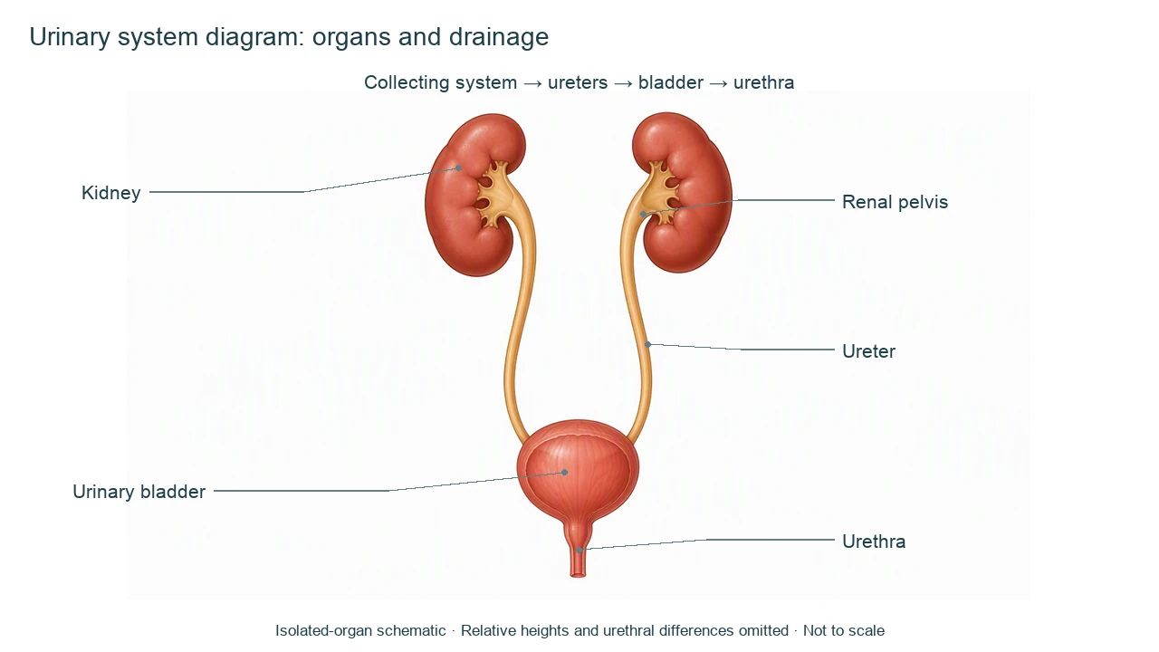
Urinary System Diagram label key
Use this label key to plan your diagram. Soft tissues or functions described in the table may require an additional view or layer.
| Label or stage | Position or relationship | Meaning and drawing check |
|---|---|---|
| Kidneys | Paired retroperitoneal organs | Produce urine through filtration and tubular processing; also contribute to endocrine and homeostatic functions. |
| Renal pelvis | Collecting region at each kidney hilum | Receives urine from calyces and continues into the ureter; not a blood vessel. |
| Ureters | One tube from each kidney to bladder | Muscular tubes propel urine toward the bladder. They are not the urethra. |
| Urinary bladder | Pelvic reservoir | Stores urine before voiding; it does not make urine. |
| Urethra | Outlet from bladder | Carries urine outside the body. Length and surrounding anatomy differ between anatomical models. |
| Urine pathway | Kidney collecting system → ureter → bladder → urethra | Follow the connections; the diagram omits microscopic nephrons and renal vessels. |
Anatomical relationships: OpenStax: Gross Anatomy of Urine Transport. On small screens, scroll the table horizontally.
Read the urinary system diagram in context
A urinary system diagram explains organ connections. Its drainage route, microscopic transport and vascular supply are related but distinct views.
01
Follow urine, not blood
The urinary system diagram shows drainage. Renal arteries and veins require a distinct vascular panel or legend.
02
Separate two scales
A urinary system diagram locates organs; a kidney section shows internal regions; a nephron diagram explains microscopic transport.
03
State anatomical scope
A simplified isolated-organ view does not show all pelvic relationships, urethral differences or the usual slightly lower right kidney.
Common urinary system diagram mistakes
Confusing ureter with urethra
Two ureters reach the bladder; the urethra leaves it. Use full words on a urinary system diagram.
Making the bladder produce urine
The kidneys produce urine, while the bladder stores it.
Sending urine through a renal vein
Veins return blood to circulation. Urine leaves through the collecting system and ureter.
Create your own urinary system diagram
Start with the prefilled prompt in the generator above. Reading the reference is free; creating a new AI draft requires an account and available credits. See current pricing.
Choose Science Drawing for an illustrated draft, or Editable Diagram when you need separate shapes and text labels. Check the anatomy in either mode before using the result.
- 1
Set the scope
Choose the view, audience and labels for your urinary system diagram. Keep advanced detail in separate panels.
- 2
Generate a focused draft
Use the prompt as a starting point. Separate labels, structures and arrows so the diagram remains readable.
- 3
Review before sharing
Check every label, connection and model assumption against the sources. Keep an answer key for a blank worksheet.
A prompt you can adapt
Create a urinary system diagram as a frontal isolated-organ schematic. Show two kidneys, their collecting pelvises, two ureters entering the bladder and one urethra leaving it. Add a separate urine flow key: collecting system to ureter to bladder to urethra. Do not use red and blue vessels as urine paths. State not to scale and that urethral/pelvic differences are omitted. Keep labels editable for checking.
Use this prompt in the generatorUrinary system FAQ
What does a urinary system diagram label?
Kidneys, ureters, bladder and urethra are the core organs; add renal pelvis for the collecting connection.
What is the order of urine flow?
After formation and collection within the kidneys, urine passes through ureters to the bladder and exits through the urethra.
What is the difference between ureter and urethra?
A ureter connects a kidney to the bladder. The urethra connects the bladder to the exterior.
Does the bladder filter blood?
No. The kidneys carry out filtration and tubular processing. The bladder stores urine.
Is this a nephron diagram?
No. A nephron diagram shows microscopic filtration, reabsorption and secretion within the kidney.
Is urinary anatomy identical in everyone?
No. Urethral length, pelvic relationships and variations require a specified model or more detailed view.
Can I generate a labeled or blank urinary system diagram?
Yes. Review the labeled draft first, then prepare numbered pointers and a separate key. New generation requires an account and credits.
Sources and editorial notes
- OpenStax, Anatomy and Physiology: Gross Anatomy of Urine Transport — factual background for the accompanying explanation.
- OpenStax: Gross Anatomy of the Kidney — additional educational context.
Prepared by the AcaDraw product team. Source and label check: . Independent medical review has not been completed. This material is for education; AI drafts need review against reliable anatomy references before use and are not for diagnosis or treatment.
Turn the reference into your own teaching figure
Choose the structures your lesson needs, create a draft, and review it before sharing.
Read the urinary system diagram guideSkin Diagram
Explore the labeled reference and drawing guide.
Endocrine System Diagram
Explore the labeled reference and drawing guide.
Lymphatic System Diagram
Explore the labeled reference and drawing guide.
Muscular System Diagram
Explore the labeled reference and drawing guide.
Kidney diagram
Explore the labeled reference and drawing guide.
Nephron diagram
Explore the labeled reference and drawing guide.
Stomach diagram
Explore the labeled reference and drawing guide.
Female reproductive system diagram
Explore the labeled reference and drawing guide.
Lungs diagram
Explore the labeled reference and drawing guide.
Nervous system diagram
Explore the labeled reference and drawing guide.
Labeled heart diagram
Explore the labeled reference and drawing guide.
Foot Anatomy diagram
Explore the labeled reference and drawing guide.
Shoulder Anatomy diagram
Explore the labeled reference and drawing guide.
Labeled brain diagram
Explore the labeled reference and drawing guide.
Digestive system diagram
Explore the labeled reference and drawing guide.
Respiratory system diagram
Explore the labeled reference and drawing guide.
Skeletal system diagram
Explore the labeled reference and drawing guide.
Neuron diagram
Explore the labeled reference and drawing guide.
Medical illustration generator
Plan other educational anatomy illustrations.