Foot Anatomy Diagram Generator
Explore foot anatomy from the top and both sides. Identify the bones, understand the arches and joints, and plan a clear diagram of the tendons and soft tissues.
SEE ACADRAW IN ACTION
Science drawing, from idea to editable figure
Create a science illustration with AI. Refine drawings, build editable diagrams, and convert images to SVG.
Demos play as you scroll into view. Choose a feature to explore.
Trusted by students & researchers from 480+ universities
A global community spanning 60+ countries and regions, from QS top-ranked universities to leading research institutes

























































































University logos belong to their respective institutions and are shown solely to illustrate our user community. They do not imply any partnership or endorsement.
Tool overview
Explore the use cases, workflow, and key things to know.
26 principal bones
Group seven tarsals, five metatarsals and fourteen phalanges.
Three useful views
Choose top, medial or lateral views to make the intended structures visible.
Layered teaching
Keep bone anatomy separate from tendon, ligament and nerve overlays.
Labeled reference
Read the same foot from three views
The reference shows the right foot in superior (top), medial (big-toe side), and lateral (little-toe side) views. It is a bone illustration: muscles, tendons, plantar fascia, nerves, and blood vessels are not shown.
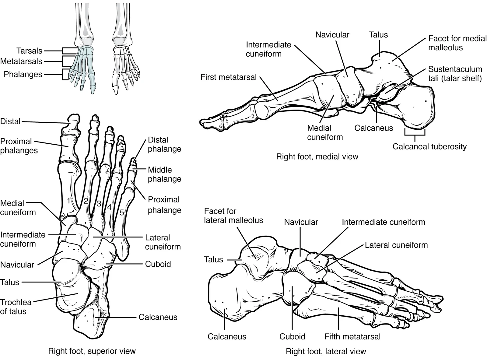
Foot anatomy labels: bones, joints and soft tissues
Use this label key alongside the reference. Soft tissues or functions described in the table may require an additional view or layer.
| Structure | Location | Role or drawing note |
|---|---|---|
| Talus | Hindfoot, above the calcaneus | Receives load from the leg at the ankle joint; it is not the heel bone. |
| Calcaneus | Heel, below the talus | Largest tarsal bone; the calcaneal (Achilles) tendon attaches posteriorly. |
| Navicular | Medial midfoot, anterior to the talus | Connects the talus to the cuneiforms and contributes to the medial arch. |
| Cuboid and three cuneiforms | Distal row of tarsal bones | Cuboid lies laterally; medial, intermediate and lateral cuneiforms lie toward metatarsals I–III. |
| Five metatarsals | Between the tarsals and toes | Number I–V from the big toe toward the little toe. |
| Fourteen phalanges | Toes | The big toe has two; each other toe usually has three. Small accessory and sesamoid bones are not included in the principal count of 26. |
| Talocrural and subtalar joints | Ankle and between talus/calcaneus | The ankle mainly allows dorsiflexion and plantar flexion; inversion and eversion involve subtalar and other intertarsal joints. |
| Achilles tendon and plantar fascia | Back of heel and sole, respectively | The tendon transmits calf muscle force to the heel. The plantar fascia helps support the longitudinal arch; it is not a tendon. |
| Extrinsic and intrinsic muscles | From the leg into the foot, or entirely within the foot | Extrinsic tendons move the ankle and toes; intrinsic muscles help control the toes and support the arches. |
Anatomical relationships: OpenStax: Bones of the lower limb. On small screens, scroll the table horizontally.
Organize a foot anatomy diagram by region
Regional names do not match the three bone groups one for one. A clear study drawing explains both systems.
01
Hindfoot: talus and calcaneus
Use a side view to show the talus above the calcaneus and the heel behind the ankle. The Achilles tendon approaches the back of the calcaneus, not the talus.
02
Midfoot: navicular, cuboid and cuneiforms
These five tarsals help form the arches and connect the hindfoot to the metatarsals. The longitudinal arches are easier to read from the side; a top view clarifies medial-to-lateral bone order.
03
Forefoot: metatarsals and phalanges
Number metatarsals I–V from the big toe. Add toe joints only when needed. Keep a separate plantar view for the plantar fascia or intrinsic muscle layers instead of inventing structures on a bone-only image.
Common foot anatomy diagram mistakes
Mixing up the side or surface
Write right/left foot and superior/plantar/medial/lateral view explicitly. The big-toe side is medial.
Counting every toe as three bones
The hallux has proximal and distal phalanges, with no middle phalanx. The usual principal bone count excludes variable accessory bones.
Using a bone drawing as a pain diagnosis
Pain can involve joints, nerves, tendons and other tissues. A diagram cannot identify the cause of an individual symptom.
Create your own foot anatomy diagram
Start with the prefilled prompt in the generator above. Reading the reference is free; creating a new AI draft requires an account and available credits. See current pricing.
Choose Science Drawing for an illustrated draft, or Editable Diagram when you need separate shapes and text labels. Check the anatomy in either mode before using the result.
- 1
Select one view and layer
Choose superior for bone order, medial for the arch, or plantar for the sole. Decide whether this is a skeleton or a soft-tissue lesson.
- 2
Generate with explicit landmarks
Specify anatomical side, view, bone groups and required tendon attachments. Use separate panels when structures overlap.
- 3
Review the count and connections
Check the hallux, metatarsal numbering, hindfoot relationships and label endpoints. Simplify the draft before exporting.
A prompt you can adapt
Create an educational labeled diagram of the right foot skeleton with a superior view and a medial side view on a white background. Mark view and anatomical side. Label talus, calcaneus, navicular, cuboid, medial/intermediate/lateral cuneiforms, metatarsals I–V, proximal/middle/distal phalanges; show only two phalanges in the hallux. In a separate side panel show Achilles tendon attaching to posterior calcaneus and plantar fascia along the sole. Keep bones and soft tissues visually distinct, use readable editable labels and uncrossed leader lines. No diagnosis or injury markers.
Use this prompt in the generatorFoot Anatomy FAQ
How many bones are in a foot?
A typical adult foot has 26 principal bones: seven tarsals, five metatarsals and fourteen phalanges. Sesamoid and accessory bones are discussed separately.
What is the top of the foot called?
The top surface is the dorsum; a top-down drawing is a dorsal or superior view. The sole is the plantar surface.
What are the three arches of the foot?
The medial longitudinal, lateral longitudinal and transverse arches. Their bony arrangement is supported by ligaments, plantar fascia and muscular activity.
Is the Achilles tendon part of the foot?
It connects the calf muscles to the posterior calcaneus at the heel. In a foot-and-ankle diagram, show that attachment in a side view.
Can this diagram explain my foot pain?
It can help you learn names and locations, but cannot diagnose pain or guide treatment. A clinician assesses symptoms, examination findings and imaging when needed.
Can I make a foot anatomy worksheet with AcaDraw?
Yes: start from the suggested prompt, review the generated draft, then remove labels to create a practice sheet. Generation requires an account and credits. Keep a checked answer key separately.
Sources and editorial notes
- OpenStax, Anatomy and Physiology: Bones of the lower limb — primary anatomy reference and illustration source.
- AAOS OrthoInfo: Foot and Ankle Conditioning Program — additional educational context.
Prepared by the AcaDraw product team. Source and label check: . Independent medical review has not been completed. This material is for education; AI drafts need review against reliable anatomy references before use and are not for diagnosis or treatment.
Turn the reference into your own teaching figure
Choose the structures your lesson needs, create a draft, and review it before sharing.
Read the step-by-step tutorial