Female Reproductive System Diagram Generator
Create a female reproductive system diagram with clear organ labels. Locate the ovaries, uterine tubes, uterus, cervix and vagina, then build a focused educational draft that keeps each structure distinct.
SEE ACADRAW IN ACTION
Science drawing, from idea to editable figure
Create a science illustration with AI. Refine drawings, build editable diagrams, and convert images to SVG.
Demos play as you scroll into view. Choose a feature to explore.
Trusted by students & researchers from 480+ universities
A global community spanning 60+ countries and regions, from QS top-ranked universities to leading research institutes
























University logos belong to their respective institutions and are shown solely to illustrate our user community. They do not imply any partnership or endorsement.
Tool overview
Explore the use cases, workflow, and key things to know.
Locate internal organs
Read the female reproductive system diagram from paired ovaries and tubes to the central uterus, cervix and vagina.
Keep the openings separate
Distinguish the vaginal canal from the urethra, which belongs to the urinary system and is not drawn here.
Specify one useful view
Use an anterior cutaway for paired structures or request a separate sagittal view for pelvic relationships.
Labeled reference
Female reproductive system diagram: an internal organ map
This female reproductive system diagram uses a simplified anterior cutaway. The ovaries sit beside the open ends of the uterine tubes; they are not sealed directly onto them. The section through the uterus distinguishes its cavity and lining from the muscular wall. External genital structures, supporting ligaments, vessels and neighboring urinary organs are omitted.
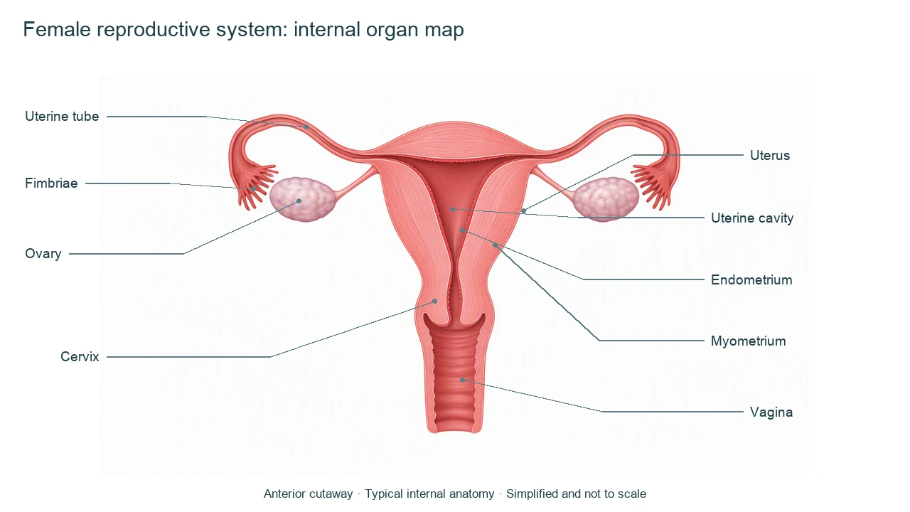
Female reproductive system diagram labels and roles
Use this label key to plan your diagram. Soft tissues or functions described in the table may require an additional view or layer.
| Structure | Where to locate it | Function and diagram note |
|---|---|---|
| Ovaries | Paired organs lateral to the uterus | Contain follicles with developing oocytes and produce reproductive hormones. Do not draw them as continuous with the tube lumen. |
| Uterine / fallopian tubes | From the upper uterine corners toward the ovaries | Help transport the oocyte toward the uterus. Fertilization usually occurs in the ampulla, a region of the tube. |
| Fimbriae | Fringed projections near each ovary | Help guide the released oocyte toward the tube opening; a simplified drawing should preserve the gap beside the ovary. |
| Uterus | Central muscular organ between the tubes and cervix | Contains the cavity where implantation normally occurs. Its shape in a frontal diagram is simplified. |
| Endometrium | Inner uterine lining | Changes during the uterine cycle; distinguish it from the thicker muscular myometrium. |
| Myometrium | Muscular layer of the uterine wall | Produces uterine contractions. It is not the hollow space at the center of the uterus. |
| Cervix | Lower narrow part of the uterus | Its canal connects the uterine cavity with the vagina. It is part of the uterus, not a separate gland. |
| Vagina | Muscular canal below the cervix | Connects the cervix with the exterior. It is distinct from the vulva and from the urinary urethra. |
Anatomical relationships: OpenStax: Anatomy and Physiology of the Female Reproductive System. On small screens, scroll the table horizontally.
Read connections in a female reproductive system diagram
A female reproductive system diagram should explain location before adding movement arrows. Different processes use the same organs in different ways.
01
Ovary to uterine tube
Ovulation releases an oocyte near the open end of a uterine tube. Fimbriae and fluid movement help bring it into the tube; the ovary does not discharge into a sealed connecting pipe.
02
Tube to uterine cavity
Ciliary activity and smooth muscle contribute to transport. Fertilization and implantation are different events: the usual fertilization site is the tubal ampulla, while normal implantation is in the uterine endometrium.
03
Uterus through cervix to vagina
The cervical canal links the cavity with the vagina. If illustrating menstrual flow, use a clearly named outward pathway rather than presenting the same arrows as the route of an oocyte.
Female reproductive system diagram errors to avoid
Joining each ovary to the tube
Leave the open relationship visible. Ovarian supporting ligaments are not the tube lumen.
Calling the entire region the vagina
Use vagina for the internal canal and vulva for external structures. This reference shows the canal, not a complete external view.
Mixing front and side views
An anterior view emphasizes paired organs. A sagittal view can show bladder and rectum relationships; make it a separate panel.
Create your own female reproductive system diagram
Start with the prefilled prompt in the generator above. Reading the reference is free; creating a new AI draft requires an account and available credits. See current pricing.
Choose Science Drawing for an illustrated draft, or Editable Diagram when you need separate shapes and text labels. Check the anatomy in either mode before using the result.
- 1
Choose the teaching scope
Ask for an internal female reproductive system diagram and name the view, organ labels and omitted structures.
- 2
Generate a focused draft
Use Science Drawing for a cutaway illustration or Editable Diagram for a simplified organ relationship map.
- 3
Check continuity and labels
Review the ovary–tube gap, uterine wall layers and cervix–vagina connection before sharing the figure.
A prompt you can adapt
Create a labeled female reproductive system diagram for education, using one simplified anterior cutaway of the internal organs. Show paired ovaries beside open fimbrial ends of the uterine tubes, with a visible gap rather than a sealed ovary-to-tube connection. Label ovaries, uterine tubes, fimbriae, uterus, uterine cavity, endometrium, myometrium, cervix and vagina. Keep the cervical canal continuous with the uterine cavity and vaginal canal. Omit external genitalia, pregnancy, urinary organs and vessels. Use clear leader lines, a white background and a not-to-scale caption. Do not add clinical diagnoses.
Use this prompt in the generatorFemale reproductive system FAQ
What does a female reproductive system diagram label?
An introductory internal female reproductive system diagram labels ovaries, uterine tubes, uterus, cervix and vagina. A cutaway can also identify the endometrium, myometrium and uterine cavity.
Are the ovaries directly connected to the fallopian tubes?
No. The fimbrial end lies near the ovary but is open to the peritoneal cavity. Ovarian supporting structures should not be confused with a sealed tube connection.
What is the difference between uterus and cervix?
The cervix is the lower narrow portion of the uterus. Its canal connects the uterine cavity to the vagina.
Are the vagina and vulva the same?
No. The vagina is an internal muscular canal. Vulva refers to the external genital structures, which are omitted from this reference.
Where do fertilization and implantation usually occur?
Fertilization usually occurs in the ampulla of a uterine tube. Normal implantation occurs in the uterine endometrium; these are different events and locations.
Can I make an editable female reproductive system diagram?
Yes. Choose Editable Diagram for separate shapes and labels, or Science Drawing for an illustrated draft. Generation requires an account and credits; check the result against reliable anatomy references.
Is this female reproductive system diagram medical advice?
No. It is a simplified educational model, not a diagnostic image or a treatment guide. Individual anatomy varies, and independent medical review has not been completed.
Sources and editorial notes
- OpenStax, Anatomy and Physiology: Anatomy and Physiology of the Female Reproductive System — factual background for the accompanying explanation.
- OpenStax: Fertilization — additional educational context.
Prepared by the AcaDraw product team. Source and label check: . Independent medical review has not been completed. This material is for education; AI drafts need review against reliable anatomy references before use and are not for diagnosis or treatment.
Turn the reference into your own teaching figure
Choose the structures your lesson needs, create a draft, and review it before sharing.
Read the organ labels and connections guideLungs diagram
Explore the labeled reference and drawing guide.
Nervous system diagram
Explore the labeled reference and drawing guide.
Labeled heart diagram
Explore the labeled reference and drawing guide.
Foot Anatomy diagram
Explore the labeled reference and drawing guide.
Shoulder Anatomy diagram
Explore the labeled reference and drawing guide.
Labeled brain diagram
Explore the labeled reference and drawing guide.
Digestive system diagram
Explore the labeled reference and drawing guide.
Respiratory system diagram
Explore the labeled reference and drawing guide.
Skeletal system diagram
Explore the labeled reference and drawing guide.
Neuron diagram
Explore the labeled reference and drawing guide.
Medical illustration generator
Plan other educational anatomy illustrations.