
Skeletal System Diagram: Every Major Bone, Labeled and Explained
Explore a labeled skeletal system diagram of major bones, axial and appendicular groups, and the adult 206-bone count. Free blank worksheet to print.
The Skeleton at a Glance
A skeletal system diagram becomes easier to read when you separate the body's central framework from its limbs. The axial skeleton includes the skull, vertebral column and thoracic cage. The appendicular skeleton includes the shoulder and hip girdles plus the upper and lower limbs.
A typical adult skeleton contains 206 bones: 80 axial and 126 appendicular. That is a counting convention for mature bones, not a promise that a whole-body image can display every small bone separately. This guide labels the major bones and groups, then accounts for the full count in a separate table.
For classroom practice, download the free blank skeletal system worksheet and answer key (two-page A4 PDF). It is available without an account or credits. To customize the labels or layout, open the skeletal system diagram generator.
Labeled Skeletal System Diagram: Front and Back Views

AI-generated educational illustration, with labels and leader lines added by AcaDraw. It shows major bone shapes and groups, not every individual adult bone. Simplified and not to scale; the accompanying explanations provide the classification and count.
The anterior view faces you. The person's right side therefore appears on your left. The posterior view shows the back, making the scapulae and spinal column easier to follow. Anatomical position means standing upright with palms facing forward; radius and ulna labels depend on this convention.
Read the leader line all the way to its endpoint. A label beside the pelvis may indicate a hip bone, the sacrum or the vertebral column. Proximity alone does not identify the structure.
Major Bones, Locations and Functions
| Bone or group | Location | Main function or identifying feature |
|---|---|---|
| Cranial bones | Upper and posterior skull | Enclose and protect the brain; eight bones in the standard adult count. |
| Facial bones | Face and jaws | Support facial structure, the nasal and oral cavities, and tooth-bearing regions; fourteen bones. |
| Vertebral column | Neck and back | Supports the trunk, protects the spinal cord and allows controlled trunk movement. |
| Ribs | Twelve pairs around the chest | Help protect thoracic organs and move with breathing. |
| Sternum | Midline front of the chest | Connects with clavicles and rib cartilages; forms the anterior thoracic cage. |
| Clavicle | Between sternum and scapula | Acts as a strut supporting shoulder position. |
| Scapula | Posterior shoulder | Provides muscle attachments and the socket that articulates with the humerus. |
| Humerus | Upper arm | Articulates at the shoulder and elbow; provides muscle attachment sites. |
| Radius | Thumb side of forearm | Contributes strongly to the wrist joint and rotates around the ulna during forearm rotation. |
| Ulna | Little-finger side of forearm | Forms the prominent elbow region and provides forearm stability. |
| Carpals, metacarpals, phalanges | Wrist, palm, fingers | Eight carpals, five metacarpals and fourteen phalanges in each hand. |
| Hip bones | Left and right pelvic girdle | Transfer loads between trunk and lower limbs and provide muscle attachments. |
| Femur | Thigh | Transmits body weight between hip and knee. |
| Patella | Front of knee | Lies in the quadriceps tendon and improves the muscle's leverage. |
| Tibia | Medial lower leg | Main weight-bearing bone of the lower leg. |
| Fibula | Lateral lower leg | Provides muscle attachments and contributes to ankle stability. |
| Tarsals, metatarsals, phalanges | Ankle region, foot, toes | Seven tarsals, five metatarsals and fourteen phalanges in each foot. |
A whole-body skeletal system diagram groups small bones to keep labels readable. Use the foot anatomy reference for individual tarsals and the shoulder anatomy reference for joint relationships.
Where the 206-Bone Count Comes From
The following accounting includes bones that are too small or obscured to label separately in the overview. Counts include both sides of the body.
| Division | Bones or group | Adult count |
|---|---|---|
| Axial | Skull: 8 cranial + 14 facial | 22 |
| Axial | Auditory ossicles: 3 in each ear | 6 |
| Axial | Hyoid | 1 |
| Axial | Vertebral column: 24 vertebrae + sacrum + coccyx | 26 |
| Axial | Ribs | 24 |
| Axial | Sternum | 1 |
| Axial subtotal | 80 | |
| Appendicular | Pectoral girdles: 2 clavicles + 2 scapulae | 4 |
| Appendicular | Upper limbs, including hands | 60 |
| Appendicular | Pelvic girdle: 2 hip bones | 2 |
| Appendicular | Lower limbs, including feet | 60 |
| Appendicular subtotal | 126 | |
| Total | 80 + 126 | 206 |
Each upper limb contributes 30 bones: one humerus, one radius, one ulna, eight carpals, five metacarpals and fourteen phalanges. Each lower limb also contributes 30: one femur, one patella, one tibia, one fibula, seven tarsals, five metatarsals and fourteen phalanges.
The count treats fused adult structures as single bones. Age, fusion patterns and accessory bones can change an individual's count. A child's developing skeleton should not simply be counted using this adult table.
Why the Pelvis Belongs to Two Divisions
The bony pelvis contains both axial and appendicular components. The sacrum and coccyx are axial. The two hip bones are appendicular. Each mature hip bone is formed by fusion of the ilium, ischium and pubis.
This distinction is easy to lose when a diagram colors the entire pelvic region as one block. In the illustration, locate the central sacrum separately from the surrounding hip bones. “Pelvis” names a region and structural assembly; “pelvic girdle” here identifies the paired hip bones.
Bones also do more than support movement. They protect organs, store minerals and contain marrow. Blood-cell production takes place in red marrow, whose distribution changes with age. A regional bone map and a bone-tissue diagram answer different questions.
Free Printable Skeletal System Worksheet
The worksheet uses the same AI-generated artwork, with 21 numbered prompts for major bones and groups. Most pointers identify the anterior skeleton; the scapula pointer identifies the posterior view. Use the article’s tables to classify axial and appendicular bones; the exercise does not require identifying every individual skull, wrist or ankle bone.
- Download the two-page A4 worksheet PDF, including answers.
- Open the blank worksheet as a high-resolution PNG.
- Open the separate answer key PNG.
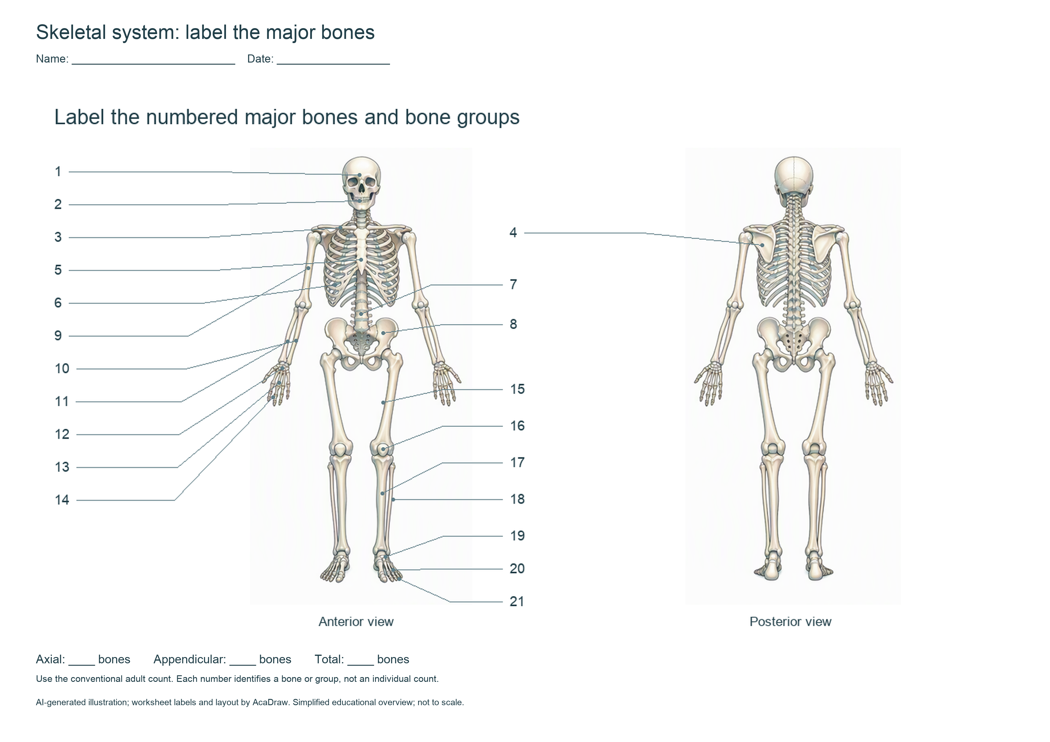
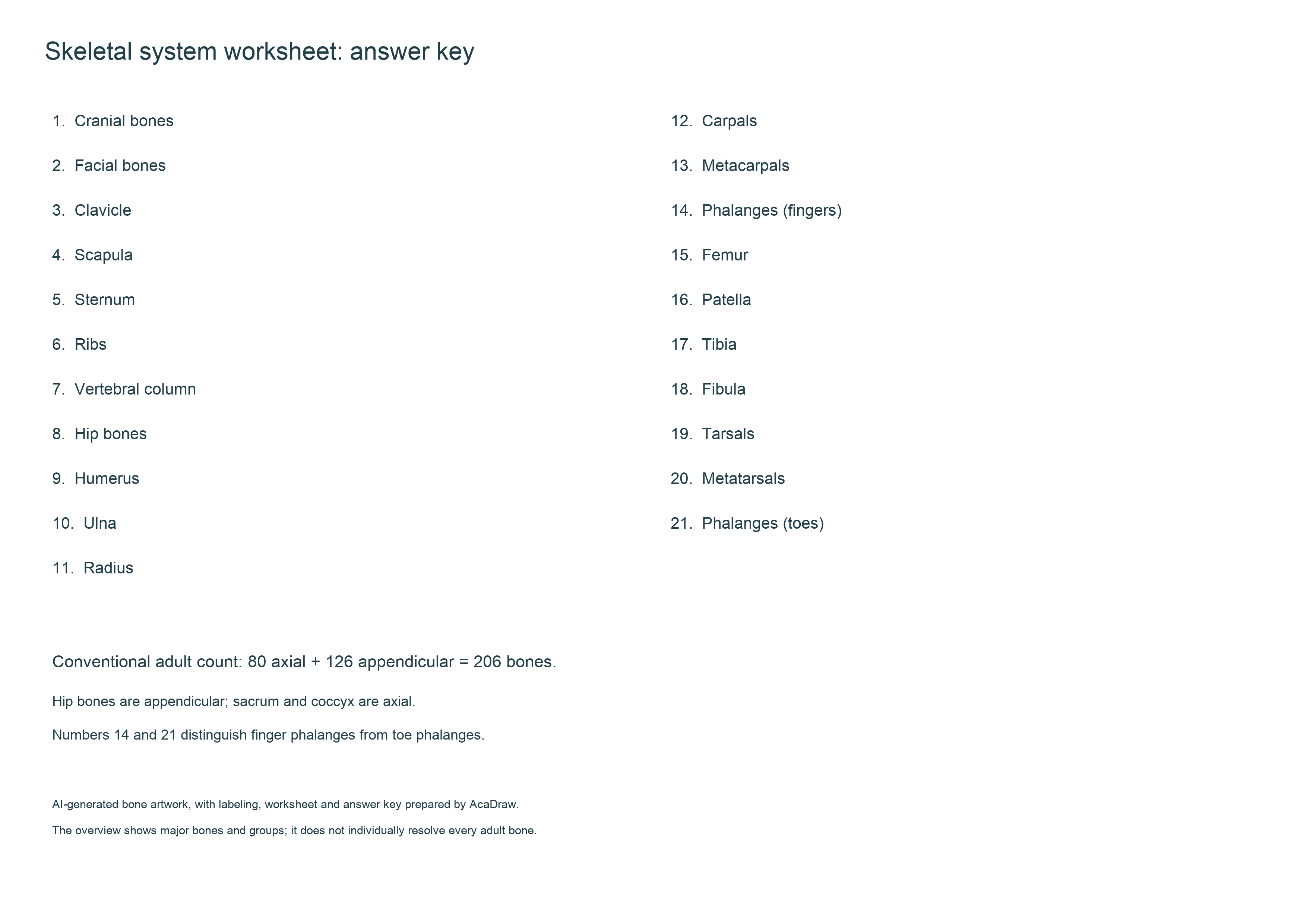
Print the A4 landscape first page for students and retain the second page as the key. Ask students to label the bones, identify an axial and an appendicular example, then complete 80 + 126 = 206. The worksheet uses AI-generated artwork with AcaDraw’s numbering and layout.
Questions That Often Cause Confusion
Are the radius and ulna reversed in a front view?
The radius stays on the person's thumb side in anatomical position. The viewer's left and right change with the view; the anatomical names do not. Check palm orientation before labeling a rotated forearm.
Is the shoulder blade part of the axial skeleton?
No. The scapula and clavicle form the pectoral girdle, part of the appendicular skeleton. Their proximity to the ribs does not change that classification.
Does this image individually label all 206 bones?
No. It labels the major bones and groups. The count table accounts for the usual 206 adult bones, while detailed regional references are needed for individual small bones.
For a version tailored to your lesson, use the skeletal system diagram generator. Request a specific view and label list, then check the generated anatomy and leader lines against a reliable reference. Generation requires an account and available credits; the worksheet downloads above are free.
References
- OpenStax Anatomy and Physiology: Divisions of the Skeletal System — adult counts and axial/appendicular divisions; cited for factual background.
- OpenStax Anatomy and Physiology: The Pectoral Girdle — clavicle and scapula.
- OpenStax Anatomy and Physiology: Bones of the Lower Limb — femur, patella, tibia, fibula and foot bones.
Prepared by the AcaDraw product team; references and diagrams checked October 23, 2026. Independent expert review has not been completed. This is an educational overview of typical adult anatomy.
Author

Categories
More Posts

Excalidraw Alternatives: 7 Tools for Diagrams & Whiteboards
Looking for an Excalidraw alternative? Compare 7 tools for precise diagrams, collaborative whiteboards, and publication-ready scientific figures.

How to Draw Lewis Dot Structures: 5 Steps
Draw any Lewis dot structure in five steps: count valence electrons, place the atoms, add bonds and lone pairs, and fix octets with multiple bonds.

AcaDraw - Create Science & Nature Level Scientific Drawings
Create Nature & Science journal-quality scientific drawings with AcaDraw's AI tool. Generate publication-ready illustrations in seconds. Try free today!
Newsletter
Join the community
Subscribe to our newsletter for the latest news and updates