Subdural Hematoma Diagram Generator
Create a subdural hematoma diagram for an introductory anatomy explanation. Locate the collection at the dura–arachnoid interface, distinguish it from epidural and subarachnoid bleeding, and review the generated labels with a reliable medical reference. This is an educational schematic, not a scan or diagnostic tool.
SEE ACADRAW IN ACTION
Science drawing, from idea to editable figure
Create a science illustration with AI. Refine drawings, build editable diagrams, and convert images to SVG.
Demos play as you scroll into view. Choose a feature to explore.
Trusted by students & researchers from 480+ universities
A global community spanning 60+ countries and regions, from QS top-ranked universities to leading research institutes























University logos belong to their respective institutions and are shown solely to illustrate our user community. They do not imply any partnership or endorsement.
Tool overview
Explore the use cases, workflow, and key things to know.
Location defines the term
Subdural, epidural and subarachnoid refer to different relationships with the meninges. Draw and label the layers before adding a collection.
Shape is only a teaching cue
A crescent is a common introductory representation. Shape alone is not sufficient for diagnosis and a generated picture is not imaging evidence.
Use a clinical reference
Actual assessment depends on symptoms, examination and medical imaging interpreted by qualified clinicians. Do not use this diagram to make care decisions.
Concept reference
Subdural Hematoma Diagram: a worked example
This exploded teaching view places the red collection below the dura and above the arachnoid. Layers are separated to make their order readable; the spacing, shape and thickness do not represent a scan or a normal cavity. The image does not establish severity, timing, mechanism or diagnosis.
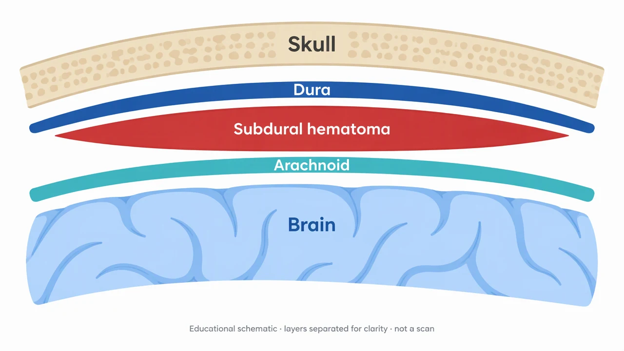
Subdural Hematoma Diagram reference key
Use the reference and definitions together. On a small screen, scroll the table horizontally.
| Element | Meaning | Review check |
|---|---|---|
| Skull | Bony boundary outside the meninges | Separate bone from the adjacent dural layer. |
| Dura mater | Outer meningeal covering | A subdural collection is internal to the dura. |
| Dura–arachnoid interface | Site of the subdural collection in the schematic | Do not present it as a large normal fluid-filled cavity. |
| Arachnoid | Meningeal layer beneath the collection | The subarachnoid space is a different compartment. |
| Brain | Neural tissue within the coverings | A schematic cannot measure pressure, midline shift or injury severity. |
Plan your subdural hematoma diagram
Identify the meningeal boundaries before interpreting the shape of a collection.
Location defines the term
Subdural, epidural and subarachnoid refer to different relationships with the meninges. Draw and label the layers before adding a collection.
Shape is only a teaching cue
A crescent is a common introductory representation. Shape alone is not sufficient for diagnosis and a generated picture is not imaging evidence.
Use a clinical reference
Actual assessment depends on symptoms, examination and medical imaging interpreted by qualified clinicians. Do not use this diagram to make care decisions.
Common mistakes to avoid
Compartment confusion
Do not place a subdural collection between skull and dura or inside the subarachnoid space.
A normal space exaggerated
The subdural region should not appear as a large normal hollow cavity.
Clinical meaning inferred from artwork
Color, width and curvature in a teaching image do not establish acuity, volume or prognosis.
Subdural Hematoma Diagram Generator: from brief to checked draft
Reading the reference is free. Generating a new draft requires an account and available credits. See current pricing.
Choose Science Drawing for an illustrated draft, or Editable Diagram for separate shapes and labels. Review all definitions, labels and connections before sharing.
- 1
Choose the teaching view
Specify the cranial section and the meningeal layers needed.
- 2
Draft and label
Generate a simplified collection with clear layer boundaries.
- 3
Review medically
Check the anatomy against a reliable reference before educational use.
A prompt you can adapt
Create a non-graphic educational subdural hematoma schematic with skull, dura, a crescent-shaped collection at the dura–arachnoid interface, arachnoid and brain labeled by short noncrossing leaders. State Schematic: not a scan, not to scale, not for diagnosis. Do not infer severity or include treatment instructions.
Use this prompt in the generatorSubdural Hematoma Diagram FAQ
Where is a subdural hematoma located?
It is a collection at the interface between the dura and arachnoid. A teaching section should distinguish it from bleeding outside the dura and within the subarachnoid space.
Is the crescent shape diagnostic?
No. It is a common schematic cue. Diagnosis requires clinical assessment and medical imaging, not a generated diagram.
How is it different from an epidural hematoma?
Epidural bleeding is outside the dura, toward the skull. Subdural bleeding is internal to the dura at the arachnoid interface.
Can I use this to assess a head injury?
No. Seek urgent medical assessment for a suspected serious head injury or new neurological symptoms. This page provides educational illustrations only.
Sources and editorial notes
Prepared by the AcaDraw product team. Source and diagram check: . This is a simplified educational model; independent expert review has not been completed. AI-generated drafts need checking against reliable references.
Explore the subdural hematoma diagram guide
Identify the meningeal boundaries before interpreting the shape of a collection.
Subdural Hematoma Diagram: Locate the Collection Between the Meninges