Immunology Diagram Generator
Use the immunology diagram generator to explain a defined interaction between immune cells and molecules. Choose the species, response type and level of detail before generating a draft. An immunology diagram can clarify selected relationships without representing the whole immune system or predicting an individual’s response.
SEE ACADRAW IN ACTION
Science drawing, from idea to editable figure
Create a science illustration with AI. Refine drawings, build editable diagrams, and convert images to SVG.
Demos play as you scroll into view. Choose a feature to explore.
Trusted by students & researchers from 480+ universities
A global community spanning 60+ countries and regions, from QS top-ranked universities to leading research institutes
























University logos belong to their respective institutions and are shown solely to illustrate our user community. They do not imply any partnership or endorsement.
Tool overview
Explore the use cases, workflow, and key things to know.
Identify the immune participants
Give each cell and molecule a precise role in the chosen interaction.
Label recognition and signaling
Distinguish peptide presentation, antigen binding and help signals.
Bound the interpretation
A selected immune interaction is not a complete immune response or a clinical prediction.
Concept reference
Immunology Diagram Generator: a worked example
The immunology diagram summarizes selected adaptive immune interactions. An antigen-presenting cell presents peptide–MHC II to a CD4 helper T cell; help signals support an antigen-activated B cell, which can differentiate into a plasma cell that secretes antibodies. The drawing omits many required contacts, co-stimulatory signals and alternative outcomes.
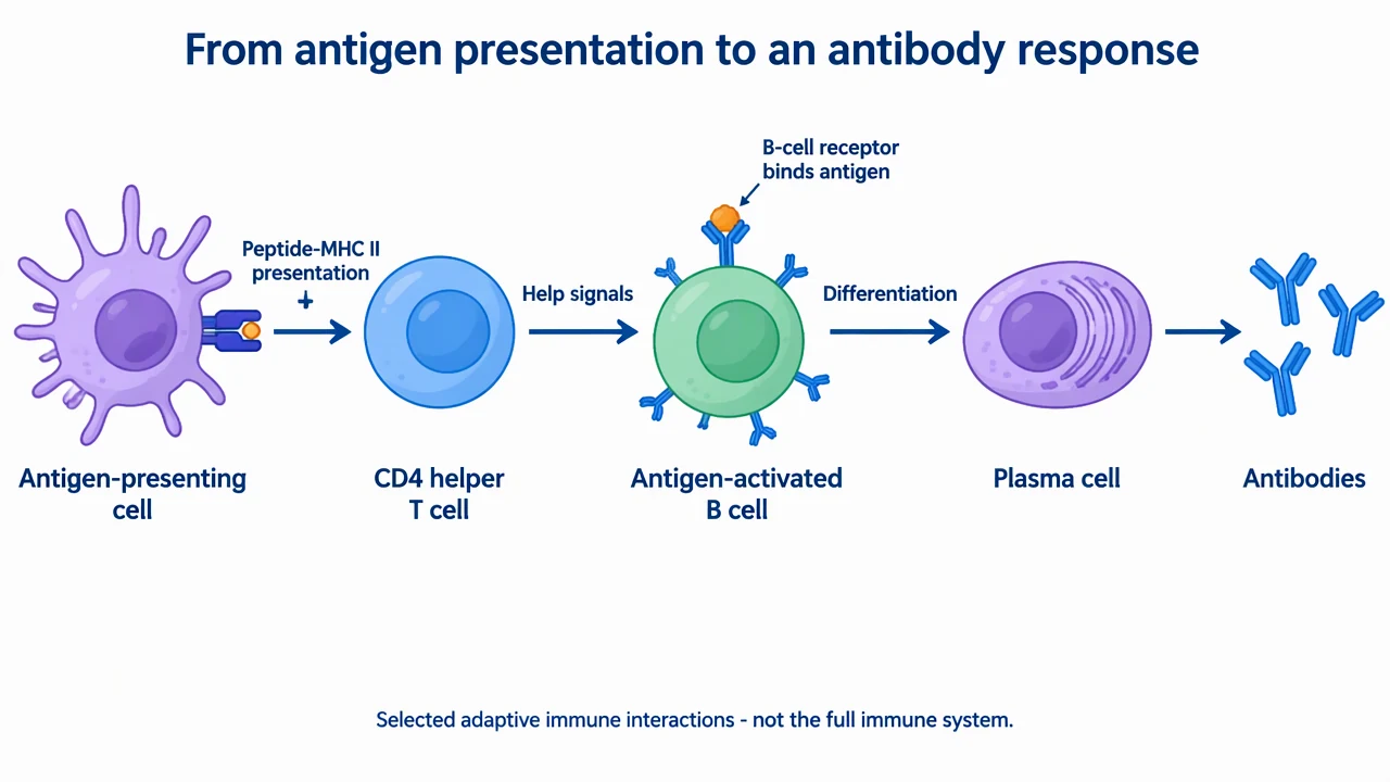
Immunology Diagram Generator: reference key
Use the reference and definitions together. On a small screen, scroll the table horizontally.
| Element | Meaning | Review check |
|---|---|---|
| Antigen-presenting cell | A cell displaying peptide–MHC complexes | Distinguish antigen processing/presentation from secretion of antibodies. |
| CD4 helper T cell | A lymphocyte receiving peptide–MHC II and additional signals | T-cell activation requires more context than a single schematic arrow. |
| B-cell receptor | A receptor that can recognize antigen | Do not confuse its binding role with T-cell recognition of peptide–MHC. |
| Help signal | Communication supporting a response | Explain omitted contact-dependent signals and cytokines in the caption. |
| Plasma cell | An antibody-secreting differentiated B-lineage cell | Place the antibody-output arrow here, not on a helper T cell. |
| Antibody | A secreted antigen-binding molecule | Do not infer protection or clinical outcome from its presence in a drawing. |
Immunology Diagram Generator: plan the content
An immunology diagram works best when cell identity, recognition and communication remain separate. Name who presents antigen, who receives the signal and which cell produces the depicted effector molecule.
Choose the response you are explaining
Separate innate recognition, antigen presentation, T-cell activation and antibody responses when one figure would otherwise become too crowded.
Keep the cell roles correct
A plasma cell secretes antibodies. A helper T cell supports selected responses through signals; it is not the source of the depicted antibodies.
Make missing steps explicit
The simplified immunology diagram omits many contacts and signals. Mention those limits instead of making one arrow look sufficient for activation.
Common mistakes in an immunology diagram
Treating the reference as the entire immune system
The drawing selects part of an adaptive response. Innate pathways, cytotoxic T cells, memory and regulatory mechanisms are largely outside its scope.
Confusing antigen recognition modes
B-cell receptors can bind antigen, whereas T-cell receptors recognize peptide presented by MHC in the relevant context. These should not be drawn as the same interaction.
Predicting protection from a schematic
A teaching immunology diagram cannot establish antibody function, protective immunity or an individual’s clinical status.
Immunology Diagram Generator: from brief to reviewed draft
Reading the reference is free. Generating a new draft requires an account and available credits. See current pricing.
Choose Science Drawing for an illustrated draft, or Editable Diagram for separate shapes and labels. Review all definitions, labels and connections before sharing.
- 1
Define one immune interaction
Tell the immunology diagram generator which cells, recognition events and outputs belong in the figure.
- 2
Draft roles and communication
Use named cells, compartment cues and labeled signals rather than a crowd of generic immune icons.
- 3
Review the immunology diagram
Check the cell functions, MHC context, arrow meanings and omitted co-signals against reliable references.
Example brief for an immunology diagram
Create an immunology diagram of selected adaptive interactions: peptide–MHC II presentation from an antigen-presenting cell to a CD4 helper T cell, help signals to an antigen-activated B cell, differentiation to a plasma cell and antibody secretion. Show the B-cell receptor binding antigen separately. State that co-signals and many immune processes are omitted and that the image makes no patient-specific claims.
Use this prompt in the generatorImmunology Diagram Generator: common questions
What does the immunology diagram generator create?
It creates an educational draft based on a specified immune process. It does not analyze patient samples or predict immunity.
Which cell secretes the antibodies in the reference?
The plasma cell, a differentiated B-lineage cell, secretes the depicted antibodies. The helper T cell is not their source.
Does an immunology diagram need every activation signal?
Not every overview must show every signal, but omitted requirements should be acknowledged so a simplified arrow does not imply sufficient activation.
Can the immunology diagram generator depict innate immunity?
Yes. Specify the relevant cells, receptors and events, and review the resulting draft rather than reusing the adaptive example unchanged.
Does the picture show clinical protection?
No. It shows selected relationships, not antibody function, a vaccine outcome or a patient-specific assessment.
Sources and editorial notes
- OpenStax: Adaptive immune response — T lymphocytes
- OpenStax: Adaptive immune response — B lymphocytes and antibodies
Prepared by the AcaDraw product team. Source and diagram check: . This is a simplified educational model; independent expert review has not been completed. AI-generated drafts need checking against reliable references.
Read the immunology diagram guide
An immunology diagram works best when cell identity, recognition and communication remain separate. Name who presents antigen, who receives the signal and which cell produces the depicted effector molecule.
Immunology Diagrams: Antigen Presentation and the B-Cell Response