Flow Cytometry Diagram Generator
Use a flow cytometry diagram generator to map cell suspension, laser interrogation, signals and gating. Explain controls and thresholds without inventing event counts or biological conclusions.
SEE ACADRAW IN ACTION
Science drawing, from idea to editable figure
Create a science illustration with AI. Refine drawings, build editable diagrams, and convert images to SVG.
Demos play as you scroll into view. Choose a feature to explore.
Trusted by students & researchers from 480+ universities
A global community spanning 60+ countries and regions, from QS top-ranked universities to leading research institutes























University logos belong to their respective institutions and are shown solely to illustrate our user community. They do not imply any partnership or endorsement.
Tool overview
Explore the use cases, workflow, and key things to know.
Define the purpose
State whether the figure explains a setup, process, interpretation or reporting boundary.
Label the convention
Explain what each arrow, line, color or control means.
Review the limits
Keep generated visuals separate from real measurements and source files.
Concept reference
Flow cytometry: from cells to a gated population
A workflow from single-cell suspension through laser interrogation and signal channels to a selected scatter-plot gate. No event counts, percentages or patient data are shown.
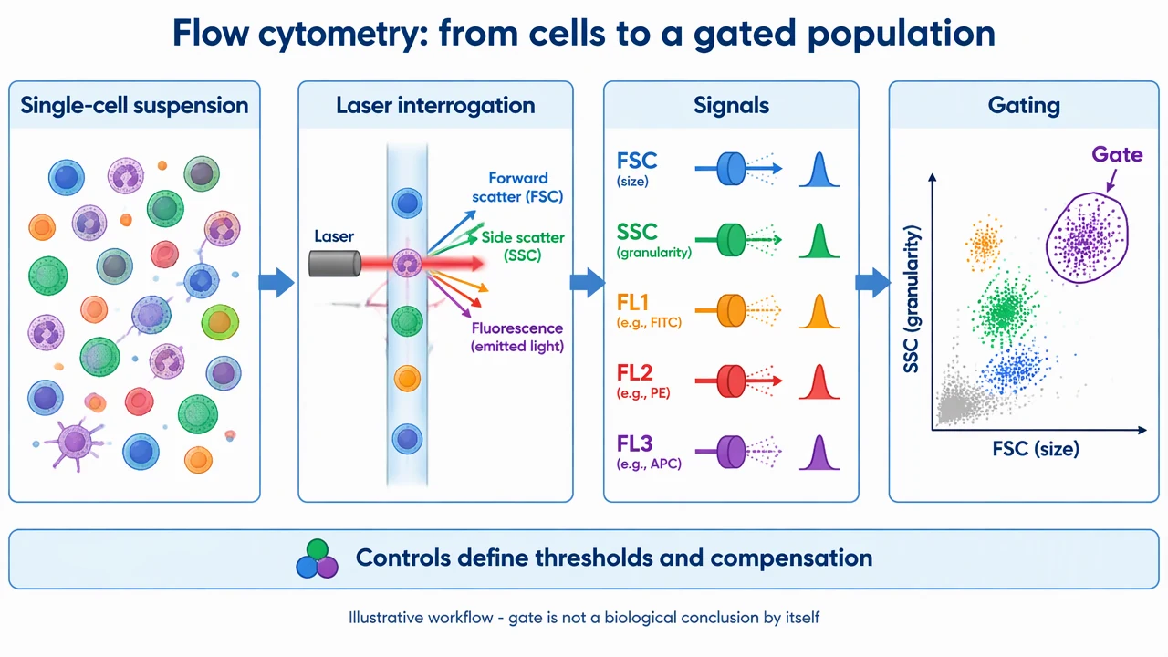
Flow Cytometry Diagram Generator: reference key
Use the reference and definitions together. On a small screen, scroll the table horizontally.
| Element | Meaning | Review check |
|---|---|---|
| FSC | A light-scatter channel | Use the instrument and sample context to interpret size-related signal. |
| SSC | A light-scatter channel | Granularity interpretations are context-dependent. |
| Fluorescence | A labeled signal channel | Name fluorophore, detector and compensation context when relevant. |
| Gate | An analysis boundary | A gate defines selected events; it does not prove cell identity. |
| Control | A threshold or reference sample | Controls support decisions about background and spillover. |
| Event | A recorded particle or cell observation | Do not add counts or percentages without data. |
Flow Cytometry Diagram Generator: plan the content
Use the flow cytometry diagram generator to connect physical measurement with an analysis decision. FSC, SSC, fluorescence channels and gates should be named separately, and a gate is not a conclusion by itself.
Start with the purpose of the flow cytometry diagram
A useful flow cytometry diagram begins with one question. Decide whether the visual should explain setup, process order, interpretation or a reporting boundary. A crowded diagram can contain accurate icons and still fail if the reader cannot tell what to notice first. The reference image is deliberately bounded. It uses labels and arrows to organize a teaching explanation, not to substitute for a protocol, a result file or a validated device. Write the scope in the caption before adding detail.
Separate the visual convention from the claim
Colors, line weights and arrows help a reader follow a flow cytometry diagram, but they do not automatically represent measured magnitude or certainty. Define what an arrow means and state when a panel is illustrative. If the figure includes a control, label what decision that control supports. This distinction matters when a familiar visual resembles a result. A band, test line, gate, signal or property label can look authoritative even when it is only a teaching placeholder. Preserve that boundary in the legend and surrounding text.
Keep provenance and review visible
Before sharing a flow cytometry diagram, compare every label with a reliable source and the actual question. Record the source image, method context, assumptions and edits. If the topic involves an assay or material, keep experimental observations in their own record rather than filling gaps with a plausible generated visual. A concise reference key makes review easier. It tells collaborators which terms are structural, which are process events and which are interpretations. That key is often more valuable than adding another decorative arrow.
Common mistakes in a flow cytometry diagram
Separate the visual convention from the claim
Colors, line weights and arrows help a reader follow a flow cytometry diagram, but they do not automatically represent measured magnitude or certainty. Define what an arrow means and state when a panel is illustrative. If the figure includes a control, label what decision that control supports. This distinction matters when a familiar visual resembles a result. A band, test line, gate, signal or property label can look authoritative even when it is only a teaching placeholder. Preserve that boundary in the legend and surrounding text.
Keep provenance and review visible
Before sharing a flow cytometry diagram, compare every label with a reliable source and the actual question. Record the source image, method context, assumptions and edits. If the topic involves an assay or material, keep experimental observations in their own record rather than filling gaps with a plausible generated visual. A concise reference key makes review easier. It tells collaborators which terms are structural, which are process events and which are interpretations. That key is often more valuable than adding another decorative arrow.
Use the draft as a reviewable starting point
The flow cytometry diagram tool helps turn a brief into a visual draft. It cannot decide whether a sample is valid, whether a mechanism is complete or whether a conclusion follows from a measurement. Ask a domain reviewer to check the final figure when the visual will guide an experiment, publication or health decision. Export the figure with a caption that names the scope and limits. Keep real data, source files and calculation records linked to the final version. A transparent workflow lets readers understand what the image explains and what it leaves unanswered.
Flow Cytometry Diagram Generator: from brief to reviewed draft
Reading the reference is free. Generating a new draft requires an account and available credits. See current pricing.
Choose Science Drawing for an illustrated draft, or Editable Diagram for separate shapes and labels. Review all definitions, labels and connections before sharing.
- 1
Write a bounded brief
Describe the scope, participants and visual conventions needed for this figure.
- 2
Generate a structured draft
Choose an illustrated or editable layout, then keep labels and arrows explicit.
- 3
Review before sharing
Compare the result with the source, method or design record and revise unsupported claims.
Example brief for a flow cytometry diagram
Original flow cytometry teaching diagram on white 1280x720 landscape, polished vector-like scientific infographic, large readable labels. Title 'Flow cytometry: from cells to a gated population'. Four panels left to right: 'Single-cell suspension' with varied cells, 'Laser interrogation' with cells crossing a laser beam and emitted light arrows, 'Signals' with FSC, SSC and fluorescence labels, 'Gating' with a simple scatter plot containing a clearly outlined selected cluster labeled 'Gate'. Add a bottom note 'Controls define thresholds and compensation'. Footer 'Illustrative workflow - gate is not a biological conclusion by itself'. No real event counts, percentages, patient data or fake statistics. Render all arrows and labels directly.
Use this prompt in the generatorFlow Cytometry Diagram Generator: common questions
What does the flow cytometry diagram tool create?
It creates an educational draft from your description. It does not infer real measurements or validate an experiment.
Does a gate identify a cell type?
Not by itself. Gating depends on markers, controls, compensation and the biological question.
What should I review before sharing?
Check labels, directions, scope and the source-specific details in the flow cytometry diagram figure.
Does it support real data analysis?
No. This page is for qualitative visual explanation and editable drafting, not computation from a data table.
Sources and editorial notes
Prepared by the AcaDraw product team. Source and diagram check: . This is a simplified educational model; independent expert review has not been completed. AI-generated drafts need checking against reliable references.
Read the flow cytometry diagram guide
A flow cytometry diagram connects physical measurement with an analysis decision. FSC, SSC, fluorescence channels and gates should be named separately, and a gate is not a conclusion by itself.
Flow Cytometry Diagrams: Gates, Controls, and Signals