Mitochondria Diagram Generator
Create a labeled mitochondria diagram that separates the outer membrane, intermembrane space, inner membrane, cristae and matrix. Use the structural reference to support a cellular-respiration lesson, then review every label and arrow.
SEE ACADRAW IN ACTION
Science drawing, from idea to editable figure
Create a science illustration with AI. Refine drawings, build editable diagrams, and convert images to SVG.
Demos play as you scroll into view. Choose a feature to explore.
Trusted by students & researchers from 480+ universities
A global community spanning 60+ countries and regions, from QS top-ranked universities to leading research institutes























University logos belong to their respective institutions and are shown solely to illustrate our user community. They do not imply any partnership or endorsement.
Tool overview
Explore the use cases, workflow, and key things to know.
Orient the two membranes
Keep the outer membrane outside and the inner membrane folded inside it.
Name spaces, not just lines
The intermembrane space lies between the two membranes; the matrix is enclosed by the folded membrane.
Connect structure to respiration
Place the Krebs cycle mainly in the matrix and oxidative phosphorylation at the inner membrane in the conventional model.
Labeled teaching reference
Mitochondria diagram: membranes, cristae, matrix, and cellular respiration context
This cutaway mitochondrion emphasizes the double-membrane organization and folded inner mitochondrial membrane. It is a generalized organelle model, not a microscopy image or a complete map of mitochondrial DNA, ribosomes, transporters and respiratory complexes.
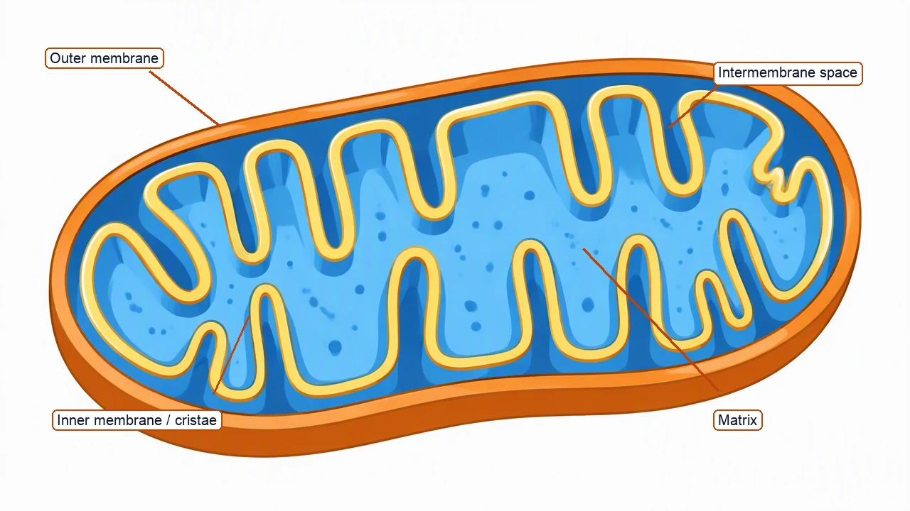
Mitochondria diagram labels and functions
Use the labels with the reference, then choose the detail level your lesson needs. On a small screen, scroll the table horizontally.
| Structure | Position in the schematic | Function or drawing check |
|---|---|---|
| Outer membrane | External boundary | Surrounds the organelle and is distinct from the selectively specialized inner membrane. |
| Intermembrane space | Between outer and inner membranes | Supports the proton-gradient model; do not draw it as the matrix. |
| Inner membrane | Folded membrane inside the organelle | Contains respiratory-chain complexes and ATP synthase in the conventional cellular-respiration model. |
| Cristae | Folds of the inner membrane | Increase inner-membrane surface area in the schematic; their exact shape varies by cell and condition. |
| Matrix | Space enclosed by the inner membrane | Contains most enzymes of the Krebs cycle and other mitochondrial components. |
| Mitochondrial genome | Within the matrix in a simplified inset | Mitochondria have their own genetic material, but this overview does not depict its copy number or nucleoids. |
Read a mitochondria diagram without overclaiming ATP
A structural mitochondria diagram shows compartments and membrane folds. It can support a respiration lesson, but it does not by itself calculate ATP yield or represent every respiratory complex.
Matrix and inner membrane have different jobs
The matrix hosts most Krebs-cycle enzymes; the folded membrane hosts the respiratory chain and ATP synthase.
Cristae connect within the membrane
Keep the cristae connected within the membrane and avoid turning each fold into a new compartment.
Cell context changes details
Mitochondria vary among tissues and organisms. A generalized diagram should say what it leaves out.
Common mitochondria diagram mistakes
Putting the Krebs cycle in the intermembrane space
Use the matrix for most Krebs-cycle enzymes in the conventional eukaryotic model.
Drawing the cristae as a smooth boundary
Cristae are folds of the inner mitochondrial membrane and are central to the structural explanation.
Treating the diagram as an ATP calculator
ATP yield depends on assumptions, shuttles and conditions; label the diagram as qualitative unless it includes a defined model.
Create your own mitochondria diagram
Reading the reference is free. Generating a new draft requires an account and available credits. See current pricing.
Choose Science Drawing for an illustrated draft, or Editable Diagram for separate shapes and text labels. Review the science and all connections before using either result.
- 1
Choose structural detail
Decide whether the figure needs only compartments, cristae detail or a respiration-location inset.
- 2
Keep spaces distinct
Trace the external boundary, intermembrane space, folded membrane and matrix in that order.
- 3
Review process placement
Check that matrix and inner-membrane labels match the intended respiration model before export.
A prompt you can adapt
Create a labeled mitochondria diagram in cutaway view. Show the outer membrane, intermembrane space, folded inner membrane with cristae, and matrix. Add a compact context inset that places most Krebs-cycle enzymes in the matrix and oxidative phosphorylation at the inner membrane. Keep the organelle clearly separate from a whole cellular-respiration pathway, use callout lines that do not cross, and do not claim the drawing is to scale or calculate ATP yield.
Use this prompt in the generatorMitochondria diagram FAQ
What are the main parts of a mitochondria diagram?
A basic diagram labels the outer membrane, intermembrane space, inner membrane, cristae and matrix. A detailed version may add mitochondrial DNA, ribosomes or respiratory complexes.
Where does the Krebs cycle occur?
Most Krebs-cycle enzymes are located in the mitochondrial matrix in the conventional eukaryotic model.
What are cristae?
Cristae are folds of the inner mitochondrial membrane. They increase surface area and contain components of oxidative phosphorylation.
Where does oxidative phosphorylation occur?
The respiratory chain and ATP synthase are associated with the inner mitochondrial membrane; proton movement is described across that membrane.
Do mitochondria have their own DNA?
Yes. Mitochondria contain their own genetic material, though a simple structural diagram may omit it or show it as a separate inset.
Can a mitochondria diagram show ATP production?
It can show the location and high-level direction of oxidative phosphorylation, but ATP yield requires a defined biochemical model and assumptions.
Sources and editorial notes
- OpenStax Biology 2e: Eukaryotic Cells
- OpenStax Biology 2e: Energy in Living Systems
- OpenStax Biology 2e: Oxidative Phosphorylation
Prepared by the AcaDraw product team. Source and diagram check: . This is an introductory educational model; independent expert review has not been completed. Generated drafts need checking against reliable references.
Create a mitochondria diagram for your lesson
Choose the scope, generate a draft, then review labels and connections before sharing.
Mitochondria Diagram: Structure, Membranes, Cristae, and Matrix