Microscope Diagram Generator
Create a microscope diagram that names the major parts of a compound light microscope and traces the simplified path from illuminator to eyepiece. Do not infer magnification, resolution or experimental results from the drawing.
SEE ACADRAW IN ACTION
Science drawing, from idea to editable figure
Create a science illustration with AI. Refine drawings, build editable diagrams, and convert images to SVG.
Demos play as you scroll into view. Choose a feature to explore.
Trusted by students & researchers from 480+ universities
A global community spanning 60+ countries and regions, from QS top-ranked universities to leading research institutes























University logos belong to their respective institutions and are shown solely to illustrate our user community. They do not imply any partnership or endorsement.
Tool overview
Explore the use cases, workflow, and key things to know.
Start at the optical path
Follow light from illuminator through condenser, specimen and objectives to the eyepiece.
Separate controls from optics
Focus knobs move the stage or optical assembly; they do not create magnification by themselves.
State the instrument scope
This is a compound light microscope overview, not a complete instrument manual.
Concept reference
Microscope diagram: labeled parts from eyepiece to base
This microscope diagram is a simplified compound light microscope. It names common components and shows a qualitative light path; exact optical layouts vary by instrument.
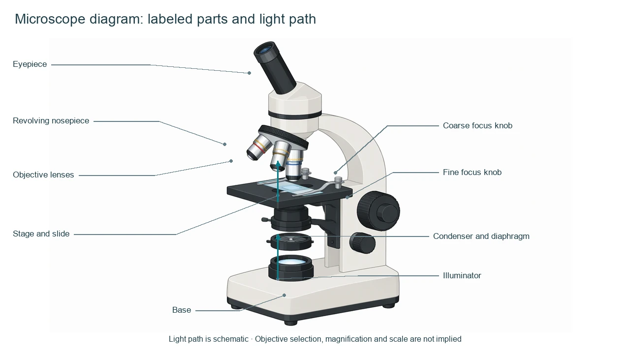
Microscope reference key
Use the reference and definitions together. On a small screen, scroll the table horizontally.
| Label or stage | Position or relationship | Meaning and drawing check |
|---|---|---|
| Eyepiece | Top viewing lens | The eyepiece is the ocular lens used by the observer. |
| Revolving nosepiece | Below the body tube | It holds and switches between objective lenses. |
| Objective lenses | Near the specimen | Objectives provide the primary magnification and must be aligned with the slide. |
| Stage and slide | Central platform | The slide rests on the stage and is held by clips or a mechanical holder. |
| Condenser and diaphragm | Below the stage | They shape and regulate illumination through the specimen. |
| Coarse and fine focus | Side controls | They bring the specimen into focus; coarse adjustment is typically used first. |
| Illuminator and base | Bottom of instrument | The illuminator supplies light and the base supports the microscope. |
Read the microscope diagram from light source to viewer
A simple path makes the instrument easier to understand: light is shaped, passes through the specimen, then enters the objective and eyepiece.
Optics form an ordered path
Keep illuminator, condenser, specimen, objective and eyepiece in sequence.
Controls change focus
Focus knobs change relative position; they are not separate optical lenses.
Real microscopes vary
Instrument design, illumination and holders differ across models.
Common microscope mistakes
Calling the stage an objective
The stage supports the slide; the objective is the lens above it.
Putting the condenser above the slide
In a transmitted-light microscope, the condenser is usually below the stage.
Claiming a magnification number
A schematic part diagram does not specify objective or eyepiece magnification.
Create your own microscope diagram
Reading the reference is free. Generating a new draft requires an account and available credits. See current pricing.
Choose Science Drawing for an illustrated draft, or Editable Diagram for separate shapes and labels. Review all definitions, labels and connections before sharing.
- 1
Choose the instrument
Specify a compound light microscope before naming its components.
- 2
Trace the path
Arrange parts so the illumination route is readable from bottom to top.
- 3
Review the mechanics
Check that the stage, objectives, condenser and focus controls are not swapped.
A prompt you can adapt
Create a labeled compound light microscope diagram. Show eyepiece, body tube, revolving nosepiece, objective lenses, stage with slide, condenser, diaphragm, coarse and fine focus controls, illuminator and base. Add a qualitative light-path arrow, keep labels editable and do not claim magnification or resolution.
Use this prompt in the generatorMicroscope diagram FAQ
What is the function of the objective lens?
The objective is the lens close to the specimen and provides the primary magnification.
What does the condenser do?
It concentrates or shapes illumination through the specimen in a transmitted-light setup.
What are coarse and fine focus for?
They adjust focus at different scales; fine focus makes smaller corrections.
Is every microscope arranged this way?
No. This is a common compound light microscope model; instruments vary.
Can I create a blank microscope worksheet?
Yes. Generate a labeled draft, then replace labels with numbered callouts and keep an answer key.
Sources and editorial notes
Prepared by the AcaDraw product team. Source and diagram check: . This is a simplified educational model; independent expert review has not been completed. AI-generated drafts need checking against reliable references.
Create a microscope diagram for your lesson
Choose the scope, generate a draft, then review labels and connections before sharing.
Microscope Diagram: Parts, Light Path, and Functions