Cell Membrane Diagram Generator
Create a cell membrane diagram with clear inside and outside labels. Explore the phospholipid bilayer, channel proteins, cholesterol and surface carbohydrates, then generate a teaching draft at the detail level you need.
SEE ACADRAW IN ACTION
Science drawing, from idea to editable figure
Create a science illustration with AI. Refine drawings, build editable diagrams, and convert images to SVG.
Demos play as you scroll into view. Choose a feature to explore.
Trusted by students & researchers from 480+ universities
A global community spanning 60+ countries and regions, from QS top-ranked universities to leading research institutes























University logos belong to their respective institutions and are shown solely to illustrate our user community. They do not imply any partnership or endorsement.
Tool overview
Explore the use cases, workflow, and key things to know.
Orient the two sides
Keep extracellular fluid above and cytosol below, with carbohydrate chains on the outer surface.
Identify structure before transport
Distinguish the lipid barrier, transmembrane channel and peripheral protein before adding any movement arrows.
Create the right membrane model
Specify an animal plasma membrane; plant, bacterial and organelle membranes need their own composition and context.
Labeled teaching reference
Cell membrane diagram: the fluid mosaic model
This cell membrane diagram shows a simplified animal plasma membrane in cross section. Hydrophilic heads face aqueous fluid on both sides; hydrophobic tails face inward. The orange carbohydrate chains project toward the extracellular side. The channel is a structural example, not a claim that every solute can pass through it.
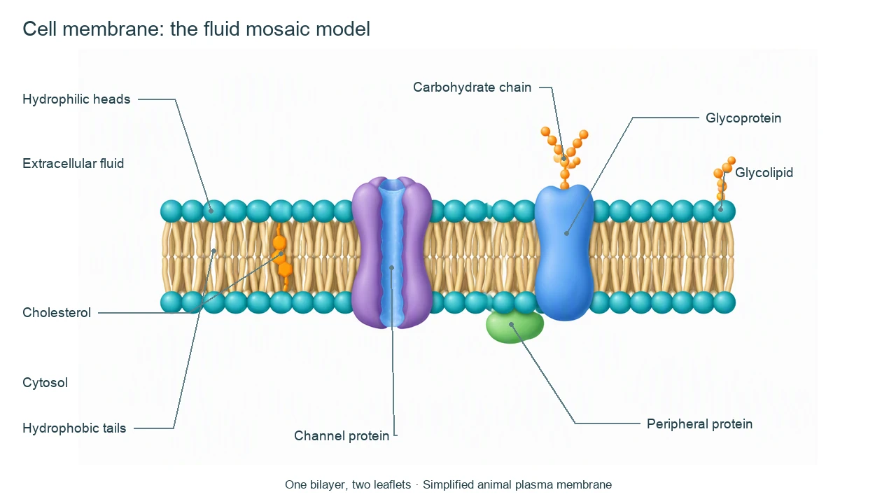
Cell membrane diagram labels and functions
Use the labels with the reference, then choose the detail level your lesson needs. On a small screen, scroll the table horizontally.
| Component | Position in this model | Main role or drawing check |
|---|---|---|
| Phospholipid heads | Both aqueous-facing surfaces | Hydrophilic head groups face extracellular fluid or cytosol, not the center of the bilayer. |
| Phospholipid tails | Hydrophobic bilayer interior | Two fatty-acid tails per phospholipid point toward the interior; the membrane has two leaflets, not two separate bilayers. |
| Channel protein | Spans the bilayer | A selective pathway through the membrane. Channel-mediated movement is passive when it follows an electrochemical gradient. |
| Integral glycoprotein | Membrane-spanning protein with an outer carbohydrate chain | The carbohydrate-bearing region faces outside the cell; specific proteins can mediate recognition, adhesion or signaling. |
| Glycolipid | Lipid with an extracellular carbohydrate chain | Part of the external cell-surface carbohydrate pattern, distinct from a glycoprotein. |
| Cholesterol | Among the lipid tails in this animal model | Modulates fluidity and permeability. It buffers temperature effects rather than always increasing or always decreasing fluidity. |
| Peripheral protein | Attached to a membrane surface | Does not span the entire bilayer. Functions depend on the protein; the illustrated inner-surface example is not every peripheral protein. |
Read structure and transport in a cell membrane diagram
A useful cell membrane diagram explains why orientation and composition matter. A cross section is a simplified snapshot of a dynamic, asymmetric surface.
Amphipathic lipids form a barrier
Each phospholipid has a hydrophilic region and hydrophobic tails. The bilayer makes a selective barrier, although it is not an absolute wall against every molecule.
The mosaic is dynamic and asymmetric
Many lipids and proteins move laterally, with constraints from interactions and the cytoskeleton. The two leaflets differ; components are not distributed as perfect mirror images.
Transport needs its own explanation
Small nonpolar molecules can cross the lipid phase. Ions generally need proteins; pumps and channels work differently. State the solute and driving force before adding arrows.
Common cell membrane diagram labeling mistakes
Putting the tails in water
Both head rows face water; the tail region is internal. Do not draw a head-to-head hydrophilic center.
Drawing carbohydrates on both sides
For the plasma membrane model, glycoprotein and glycolipid carbohydrate chains face extracellular fluid. They are not interchangeable with a cytosolic protein.
Adding ATP to every channel
A channel does not become a pump because it spans the membrane. Distinguish passive transport from ATP-driven pumping and coupled transport.
Create your own cell membrane diagram
Reading the reference is free. Generating a new draft requires an account and available credits. See current pricing.
Choose Science Drawing for an illustrated draft, or Editable Diagram for separate shapes and text labels. Review the science and all connections before using either result.
- 1
Choose the biological context
Use an animal plasma membrane or explicitly request a different membrane type and composition.
- 2
Place the bilayer and key components
Decide which proteins, carbohydrates and lipids belong in the cell membrane diagram. Mark the aqueous sides.
- 3
Review orientation and mechanism
Check heads, tails and carbohydrate placement. Add transport arrows only when the solute and energy source are specified.
A prompt you can adapt
Create a labeled cell membrane diagram of an animal plasma membrane in cross section using the fluid mosaic model. Show exactly one phospholipid bilayer: hydrophilic heads face extracellular fluid and cytosol, hydrophobic tails face the interior. Include a transmembrane channel with an aqueous pore, an integral glycoprotein and glycolipid with carbohydrate chains only on the extracellular side, cholesterol among the tails and a peripheral protein on the cytosolic face. Label inside and outside and keep leader lines separate. Do not draw a cell wall or transport arrows unless a specific solute and mechanism are requested. State that colors and proportions are schematic.
Use this prompt in the generatorCell membrane diagram FAQ
What should a cell membrane diagram label?
A basic animal cell membrane diagram labels the phospholipid bilayer, heads, tails, membrane proteins, cholesterol, carbohydrates, extracellular fluid and cytosol. Add details according to the teaching level.
What is the fluid mosaic model?
It describes a dynamic lipid bilayer containing a mosaic of proteins and other components. Many components move laterally, but their motion can be constrained and the two leaflets are asymmetric.
Which way do phospholipid heads and tails point?
Hydrophilic heads face the aqueous fluid on the outside and inside of the cell. Hydrophobic tails point toward the center of the bilayer, away from that fluid.
Where are carbohydrates in a plasma membrane diagram?
Carbohydrate chains attached to glycoproteins and glycolipids face the extracellular side. They contribute to recognition, interactions and the cell-surface glycocalyx.
Does cholesterol always make a membrane more fluid?
No. In animal membranes cholesterol buffers temperature-dependent behavior. It restrains some lipid movement at higher temperatures and helps prevent tight packing at lower temperatures.
Is a cell membrane the same as a cell wall?
No. The plasma membrane is a selective lipid-based boundary. A cell wall is an additional external structure in plants, fungi and many prokaryotes; animal cells lack cell walls.
Can I create an editable cell membrane diagram?
Yes. Use Editable Diagram for separate shapes and labels, or Science Drawing for an illustrated draft. Generation requires an account and credits. Review molecular orientation and any transport mechanism.
Sources and editorial notes
- OpenStax Biology 2e: Components and Structure
- OpenStax Biology 2e: Passive Transport
- OpenStax Biology 2e: Active Transport
Prepared by the AcaDraw product team. Source and diagram check: . This is an introductory educational model; independent expert review has not been completed. Generated drafts need checking against reliable references.
Create a cell membrane diagram for your lesson
Choose the scope, generate a draft, then review labels and connections before sharing.
Cell Membrane Diagram: Labeled Parts and the Fluid Mosaic Model