DNA Replication Diagram Generator
Create a DNA replication diagram that makes strand direction readable. Trace one replication fork, compare leading and lagging strands, and check 5′–3′ synthesis before generating your own labeled teaching draft.
SEE ACADRAW IN ACTION
Science drawing, from idea to editable figure
Create a science illustration with AI. Refine drawings, build editable diagrams, and convert images to SVG.
Demos play as you scroll into view. Choose a feature to explore.
Trusted by students & researchers from 480+ universities
A global community spanning 60+ countries and regions, from QS top-ranked universities to leading research institutes























University logos belong to their respective institutions and are shown solely to illustrate our user community. They do not imply any partnership or endorsement.
Tool overview
Explore the use cases, workflow, and key things to know.
Follow both 5′–3′ arrows
The leading and lagging strands use the same chemical direction, despite their different directions relative to the fork.
Separate strands from enzyme roles
Use a restrained fork overview, with a separate enzyme key for helicase, primase, polymerase and ligase.
Choose a biological model
Specify a general fork or a bacterial/eukaryotic lesson. Do not mix polymerase names or primer-removal systems.
Labeled teaching reference
DNA replication diagram: leading and lagging strands
This DNA replication diagram isolates one fork moving to the right. New DNA is teal and parental templates are navy. The top daughter strand extends continuously toward the fork; the lower fragments extend away from it. Both are synthesized 5′ to 3′. Orange marks RNA primers on the illustrated lagging fragments; the earlier leading-strand primer is outside this view.
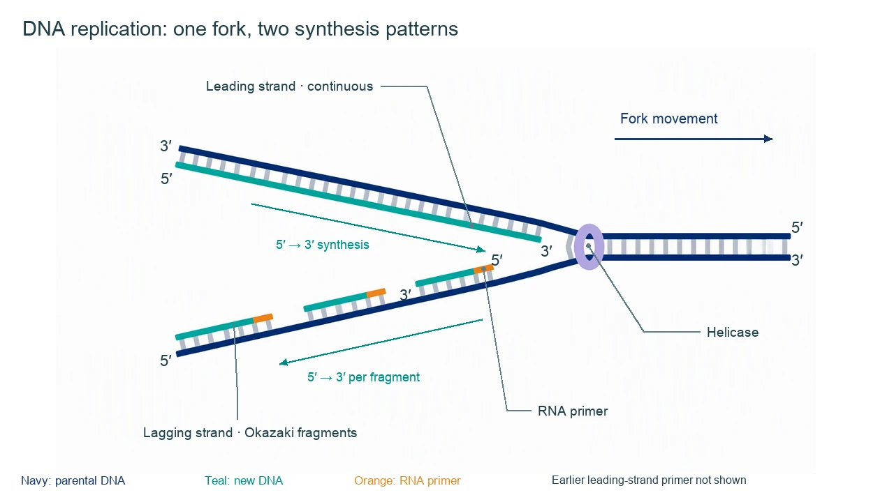
DNA replication diagram labels and enzyme roles
Use the labels with the reference, then choose the detail level your lesson needs. On a small screen, scroll the table horizontally.
| Structure or enzyme | Role at replication | What the diagram must keep clear |
|---|---|---|
| Parental templates | Guide complementary sequence | The two templates are antiparallel. Each product duplex ultimately contains one parental strand and one newly made strand. |
| Leading strand | Continuous new DNA synthesis at this fork | Its growing 3′ end progresses toward the moving fork; the earlier primer is not shown in this cropped view. |
| Lagging strand | Discontinuous synthesis as Okazaki fragments | Each fragment grows 5′ → 3′ away from the fork, starting from a new primer. Never label synthesis as 3′ → 5′. |
| Helicase | Separates parental strands | Works at the fork. It separates paired strands rather than cutting each DNA backbone into fragments. |
| Primase and RNA primers | Provide starting 3′-OH groups | The leading strand also needs an initial primer. Lagging-strand synthesis needs repeated priming. |
| DNA polymerase | Extends a primed strand | Adds nucleotides to a 3′ end while using the complementary template. Specific polymerase names depend on the organism. |
| DNA ligase | Seals remaining backbone nicks | Primer removal and DNA replacement precede joining. Ligase does not synthesize the entire missing DNA region. |
| Topoisomerase and single-strand-binding proteins | Manage torsion and exposed templates | Not drawn in this overview. Add a separate detail panel when explaining their positions or mechanism. |
Read every direction in a DNA replication diagram
A DNA replication diagram can have several arrows with different meanings. Fork movement, template orientation and new-strand extension must not be merged into a single unexplained direction.
Semiconservative describes the product
Each daughter duplex contains one parental strand and one newly synthesized strand. It does not mean that each original strand is cut in half.
Antiparallel templates explain the asymmetry
The polymerase extends only a growing 3′ end. Opposite template orientations produce one continuous strand and repeated fragments at a moving fork.
One fork is not the whole replication region
An origin usually gives rise to two forks in a bidirectional model. This image shows only the right-moving fork; it is not a complete replication bubble or chromosome.
Common DNA replication diagram direction mistakes
Making the lagging strand grow 3′ → 5′
Both new strands grow 5′ → 3′. Lagging fragments grow away from this fork, rather than using a different chemical direction.
Forgetting leading-strand priming
Continuous extension still requires an initial primer. A simplified fork close-up may omit the primer because it lies earlier along the strand.
Asking ligase to fill every gap
Primer removal and replacement create DNA between fragments. Ligase then seals nicks in the backbone; enzyme details vary by organism.
Create your own dna replication diagram
Reading the reference is free. Generating a new draft requires an account and available credits. See current pricing.
Choose Science Drawing for an illustrated draft, or Editable Diagram for separate shapes and text labels. Review the science and all connections before using either result.
- 1
Choose the fork model
Specify one right-moving fork or an entire bidirectional replication bubble, plus the organism if naming enzymes.
- 2
Set template polarity first
Mark parental 5′ and 3′ ends, then add complementary daughter strands and separate synthesis arrows.
- 3
Review primers and fragment direction
Check the growing 3′ ends, each lagging primer and the distinction between primer replacement and ligation before exporting.
A prompt you can adapt
Create a labeled DNA replication diagram showing one fork moving to the right. Use navy parental strands and teal new DNA. Set the upper template 3′ at left and 5′ at right; its new leading strand is 5′ at left with a growing 3′ end toward the fork. Set the lower template 5′ at left and 3′ at right; show separate Okazaki fragments, each with an RNA primer at its fork-proximal 5′ end and 5′ → 3′ growth away from the fork. Label helicase, fork motion and the two distinct synthesis directions. State that the leading strand also needed an earlier primer, omitted from this close-up. Put primase, polymerase, primer removal and ligase in a separate enzyme key; do not mix bacterial and eukaryotic enzyme names.
Use this prompt in the generatorDNA replication diagram FAQ
What should a DNA replication diagram show?
A useful fork diagram shows parental templates, new DNA, leading and lagging strands, primers, strand polarity and fork movement. Add an enzyme key without crowding the strand directions.
Do both DNA strands grow 5′ to 3′?
Yes. DNA polymerase extends a growing 3′ end, so each newly synthesized strand or Okazaki fragment grows 5′ to 3′. The two templates have opposite orientations.
What is the difference between leading and lagging strands?
At a given fork the leading strand is extended continuously toward fork progression. The lagging strand is made in fragments that extend away from the fork and are later joined.
Does the leading strand need an RNA primer?
Yes. Continuous synthesis still needs an initial primed 3′ end. A close-up of an established fork can omit that earlier primer, but the text should explain the omission.
What does semiconservative replication mean?
Each daughter DNA duplex contains one parental strand and one newly synthesized strand. It describes strand inheritance in the product, not a mixture of short old and new pieces within each strand.
Where does nuclear DNA replication fit in the cell cycle?
It occurs during S phase of interphase, before mitosis. Bacterial replication and division do not use the eukaryotic G1, S, G2 and M framework.
Can I generate an editable DNA replication diagram?
Yes. Editable Diagram gives separate shapes and labels; Science Drawing gives an illustrated draft. Generation needs an account and credits. Verify the strand ends, primers and arrows before use.
Sources and editorial notes
- OpenStax Biology 2e: Basics of DNA Replication
- OpenStax Biology 2e: DNA Replication in Prokaryotes
- OpenStax Biology 2e: DNA Replication in Eukaryotes
Prepared by the AcaDraw product team. Source and diagram check: . This is an introductory educational model; independent expert review has not been completed. Generated drafts need checking against reliable references.
Create a dna replication diagram for your lesson
Choose the scope, generate a draft, then review labels and connections before sharing.
DNA Replication Diagram: Fork Direction, Strands, and Enzymes