Bacterial Cell Diagram Generator
Use this labeled bacterial cell diagram to locate the nucleoid, ribosomes and cell envelope. Separate shared cell components from optional features, then create a checked teaching draft.
SEE ACADRAW IN ACTION
Science drawing, from idea to editable figure
Create a science illustration with AI. Refine drawings, build editable diagrams, and convert images to SVG.
Demos play as you scroll into view. Choose a feature to explore.
Trusted by students & researchers from 480+ universities
A global community spanning 60+ countries and regions, from QS top-ranked universities to leading research institutes























University logos belong to their respective institutions and are shown solely to illustrate our user community. They do not imply any partnership or endorsement.
Tool overview
Explore the use cases, workflow, and key things to know.
Identify the cell envelope
Read membrane, wall and capsule as different structures.
Separate core and optional parts
Do not give every bacterium a capsule, plasmid or flagellum.
Build a reviewable draft
Check endpoints and organism-specific details before export.
Labeled teaching reference
Labeled bacterial cell diagram: core and optional structures
This generalized rod-shaped cell is a cutaway schematic, not a portrait of every bacterium. It shows a cell wall outside the plasma membrane and a capsule outside the wall. An outer membrane is omitted, so the envelope is not a complete Gram-negative model. Accessory features are marked optional, and some bacteria lack a cell wall.
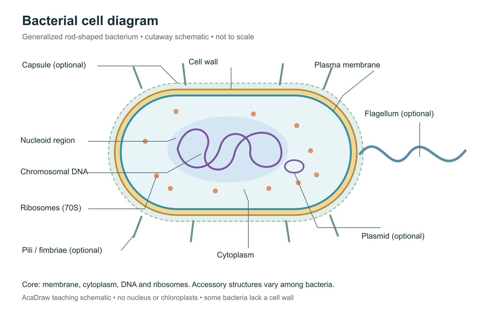
Bacterial cell diagram labels and their functions
Use the labels with the reference, then choose the detail level your lesson needs. On a small screen, scroll the table horizontally.
| Structure | Location in the schematic | Function and drawing note |
|---|---|---|
| Plasma membrane | Inner boundary of the cell envelope | Controls exchange and supports cellular processes. Keep it distinct from the wall outside it. |
| Cell wall | Outside the plasma membrane | Provides shape and protection in most bacteria. Peptidoglycan is characteristic of bacterial walls; wall-less bacteria are exceptions. |
| Capsule or slime layer | Outside the wall, when present | A glycocalyx can support attachment and protection. Not all bacteria have one; a capsule is organized, while a slime layer is looser. |
| Cytoplasm | Inside the membrane | Contains dissolved substances and cellular components. It is not an empty gap around the chromosome. |
| Nucleoid and chromosome | DNA-containing region within the cytoplasm | The nucleoid is not enclosed by a nuclear membrane. A single circular chromosome is common, with exceptions to both number and shape. |
| Ribosomes | Small particles throughout the cytoplasm | Bacterial cytoplasmic ribosomes are 70S and synthesize proteins. The drawing enlarges a few representative particles. |
| Plasmid | Small DNA molecule separate from the chromosome | Often shown as a circular loop; plasmids are not universal or essential to every bacterial cell. |
| Pili and fimbriae | Short surface appendages, when present | Many aid attachment; some specialized pili support DNA transfer or motility. Do not treat every short appendage as a conjugation pilus. |
| Flagellum | Long surface appendage, when present | Can propel a cell by rotation. Number and arrangement vary, and many bacteria do not have flagella. |
Choose the scope of your bacterial cell diagram
A bacterial cell diagram is one example of prokaryotic organization. Prokaryotes include two distinct domains, Bacteria and Archaea; a bacterial envelope should not be used as a universal archaeal model.
Bacteria and archaea
Both lack a membrane-bound nucleus. Archaeal walls do not contain bacterial peptidoglycan, and membrane chemistry differs. Label the organism group before describing its envelope.
Gram-positive and Gram-negative
A Gram-positive envelope typically has a thick peptidoglycan layer. A Gram-negative envelope has thin peptidoglycan and an outer membrane. Use a separate envelope inset when teaching this distinction.
Bacterial and eukaryotic cells
A basic bacterium has no membrane-bound nucleus, mitochondria or chloroplasts. Photosynthetic bacteria use other membrane arrangements; they do not become plant cells.
Common bacterial cell diagram mistakes
Drawing a nucleus around the DNA
Represent a nucleoid region without a nuclear envelope. Do not add mitochondria or chloroplasts.
Making accessory parts compulsory
Mark capsule, plasmids and appendages as variable. Match a species-specific drawing to a suitable source.
Confusing membrane and wall
Use separate label endpoints. Add a separate outer membrane only when the selected envelope type calls for one.
Create your own bacterial cell diagram
Reading the reference is free. Generating a new draft requires an account and available credits. See current pricing.
Choose Science Drawing for an illustrated draft, or Editable Diagram for separate shapes and text labels. Review the science and all connections before using either result.
- 1
Choose the organism and level
Start with a generalized bacterium, or name a species and envelope type. Decide whether optional structures belong in the lesson.
- 2
Group labels by location
Separate the envelope, cytoplasmic components and appendages. Keep short leader lines clear of the DNA loops.
- 3
Check and make a worksheet
Verify cell layers and optional parts. Duplicate your checked draft without labels and keep a separate answer key.
A prompt you can adapt
Create an educational bacterial cell diagram showing a generalized rod-shaped bacterium in cutaway on a white background. Label plasma membrane, cell wall, cytoplasm, nucleoid region with chromosomal DNA, and 70S ribosomes. Show a capsule, small circular plasmid, short pili or fimbriae, and a flagellum as optional features. Do not add a membrane-bound nucleus, mitochondria or chloroplasts. State that this is a generalized model, not a complete Gram-negative envelope or a universal archaeal cell. Make leader lines end on the intended layer. Keep shapes and labels separate in Editable Diagram mode for manual review.
Use this prompt in the generatorBacterial Cell diagram FAQ
What should a bacterial cell diagram label?
Start with the plasma membrane, cytoplasm, chromosome within a nucleoid and ribosomes. Most bacteria also have a cell wall. Add capsule, plasmids, pili or flagella only when they fit the organism or are clearly marked optional.
Is a bacterial cell the same as a prokaryotic cell?
A bacterium is a prokaryote, but not every prokaryote is a bacterium. Archaea form a separate domain and differ in membrane and cell-wall chemistry.
Do bacteria have a nucleus?
They have DNA but no membrane-bound nucleus. The main chromosomal DNA occupies a region called the nucleoid.
Does every bacterium have a cell wall or flagellum?
No. Most bacteria have a cell wall, but wall-less groups exist. Flagella, capsules, pili and plasmids also vary; a generalized picture should not imply they are universal.
Why are mitochondria and chloroplasts absent?
Bacteria do not contain these membrane-bound organelles. Their energy-related processes use other cellular structures, including the plasma membrane or specialized internal membranes in some groups.
Can I make a blank bacterial cell diagram?
Yes. Create and review your labeled draft, duplicate it, and replace names with numbered pointers. Check that membrane and wall endpoints remain distinct and keep a separate answer key.
Can I generate an editable bacterial cell diagram?
Yes. Use the prompt above and choose Editable Diagram for separate shapes and text. An account and available credits are required. Review the biological model and label endpoints before teaching or sharing.
Sources and editorial notes
- OpenStax Microbiology: Unique Characteristics of Prokaryotic Cells
- OpenStax Biology 2e: Prokaryotic Cells
Prepared by the AcaDraw product team. Source and diagram check: . This is an introductory educational model; independent expert review has not been completed. Generated drafts need checking against reliable references.
Create a bacterial cell diagram for your lesson
Choose the scope, generate a draft, then review labels and connections before sharing.
How to Draw a Bacterial Cell Diagram in 5 Steps